Discovering a black spot beneath your toenail can trigger immediate concern, particularly when considering the well-documented case of Bob Marley, whose subungual melanoma proved fatal. However, the vast majority of these discolourations stem from benign causes that require minimal intervention. Understanding the diverse aetiologies behind subungual pigmentation enables proper assessment and treatment decisions, preventing unnecessary anxiety whilst ensuring serious conditions receive appropriate attention.
The nail apparatus consists of the nail matrix, nail bed, and surrounding soft tissues, each susceptible to various pathological processes that can manifest as pigmentation changes. From traumatic haematomas to systemic disease manifestations, the differential diagnosis encompasses numerous conditions requiring distinct therapeutic approaches. Modern diagnostic techniques, including dermoscopy and histopathological analysis, have revolutionised our ability to differentiate between benign and malignant lesions with unprecedented accuracy.
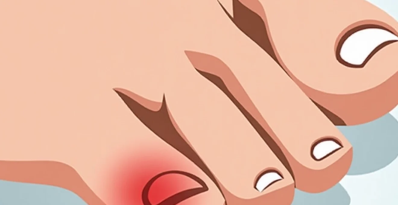
Traumatic subungual haematoma formation and acute injury mechanisms
Traumatic subungual haematomas represent the most frequent cause of black discolouration beneath toenails, accounting for approximately 85% of cases presenting to podiatry clinics. These lesions develop when mechanical forces disrupt the delicate vascular network within the nail bed, causing blood to accumulate between the nail plate and underlying tissues. The characteristic appearance typically manifests as a well-demarcated, dark red to black area that may occupy varying portions of the nail bed surface.
Blunt force trauma impact on nail bed vasculature
Direct impact injuries create immediate vascular disruption through shearing forces that tear capillaries and venules within the nail bed matrix. The confined space beneath the nail plate prevents blood dispersal, leading to progressive accumulation and characteristic discolouration. Pain intensity correlates directly with the volume of trapped blood and associated tissue pressure elevation. Most blunt force injuries occur during sporting activities, occupational accidents, or household mishaps involving heavy objects.
Repetitive microtrauma in athletes and runners
Endurance athletes frequently develop subungual haematomas through cumulative microtrauma rather than single catastrophic events. The repetitive toe-to-shoe contact during prolonged activity creates microscopic vascular damage that accumulates over time. Runner’s toe typically affects the hallux or second digit, particularly in individuals wearing poorly fitted footwear or participating in downhill running activities. The gradual onset distinguishes these lesions from acute traumatic haematomas, often presenting as slowly expanding dark areas beneath the nail.
Crushing injuries and digital compression pathophysiology
Severe crushing injuries produce extensive tissue damage extending beyond simple vascular disruption to include nail matrix and periungual soft tissue involvement. These complex injuries often result in nail loss and require surgical intervention to prevent complications such as infection or permanent deformity. The mechanism involves direct compression forces that exceed tissue tolerance limits, causing widespread cellular death and inflammatory responses. Recovery typically requires several months, with new nail growth beginning approximately six to eight weeks post-injury.
Subungual bleeding assessment using pain scale indicators
Pain assessment provides crucial diagnostic information for distinguishing acute traumatic haematomas from other causes of subungual pigmentation. Fresh haematomas typically produce severe throbbing pain due to increased pressure within the confined nail bed space. Chronic haematomas rarely cause significant discomfort, whilst malignant lesions typically remain painless throughout their development. The presence of severe pain following known trauma strongly suggests acute haematoma formation requiring potential decompression procedures.
Melanocytic lesions and subungual melanoma differentiation
Subungual melanoma represents one of the most challenging diagnostic scenarios in dermatology, accounting for approximately 2-3% of all melanoma cases but carrying significant morbidity when diagnosis is delayed. These aggressive malignancies typically arise from melanocytes within the nail matrix or proximal nail bed, manifesting as longitudinal pigmented bands or irregular dark patches. The anatomical location beneath the nail plate complicates early detection, often leading to advanced disease at presentation.
Early detection of subungual melanoma significantly improves patient outcomes, with five-year survival rates exceeding 95% when caught before metastasis occurs.
Acral lentiginous melanoma hutchinson’s sign recognition
Hutchinson’s sign represents a pathognomonic feature of subungual melanoma, characterised by pigmentation extending beyond the nail plate onto the surrounding periungual skin. This finding indicates advanced local disease with potential for rapid progression and metastatic spread. The sign typically develops gradually over months to years, distinguishing it from acute traumatic pigmentation changes. Recognition requires careful examination of the nail fold and hyponychium for subtle pigmentation extending beyond normal anatomical boundaries.
Benign melanocytic naevi versus malignant transformation
Benign melanocytic naevi can occur within the nail apparatus, presenting as stable longitudinal pigmented bands that remain unchanged over extended periods. These lesions typically demonstrate uniform pigmentation, regular borders, and consistent width throughout their length. Malignant transformation manifests as changes in pigmentation intensity, band width, or development of irregular borders within previously stable lesions. Dermoscopic examination reveals characteristic features that aid in differentiation, including regular versus irregular pigmentation patterns and the presence or absence of periungual involvement.
ABCDEF criteria application for subungual pigmentation
The modified ABCDEF criteria provide a systematic approach for evaluating suspicious subungual pigmentation, adapting traditional melanoma screening parameters for nail-specific features. Asymmetry refers to irregular pigmentation distribution, whilst Border irregularity indicates uneven band edges or extension onto periungual skin. Colour variation within the same lesion, Diameter exceeding 3 millimetres, Evolution over time, and Family history complete the assessment framework. This systematic approach improves diagnostic accuracy whilst reducing unnecessary biopsies of benign lesions.
Dermoscopic features of longitudinal melanonychia evaluation
Dermoscopic examination provides enhanced visualisation of subungual pigmentation patterns, revealing subtle features invisible to naked eye examination. Benign longitudinal melanonychia typically displays regular parallel lines with consistent spacing and uniform pigmentation throughout the band width. Malignant lesions demonstrate irregular pigmentation patterns, varying line spacing, and potential micro-Hutchinson signs visible only under magnification. Advanced dermoscopic techniques utilising polarised light improve contrast and reveal deeper pigmentation patterns within the nail matrix.
Fungal infections and Onychomycosis-Related discolouration
Fungal nail infections commonly produce secondary pigmentation changes that can mimic more serious conditions, particularly when extensive debris accumulation occurs beneath the nail plate. Onychomycosis affects approximately 10% of the global population, with increased prevalence in elderly individuals and those with compromised immune systems. The characteristic yellowing associated with fungal infections can progress to darker discolouration as secondary bacterial colonisation develops or debris accumulates within the nail bed space.
Dermatophyte infections typically begin at the distal nail edge, gradually extending proximally whilst producing characteristic nail thickening and friability. The development of black discolouration usually indicates advanced infection with significant nail destruction and potential secondary bacterial involvement. Candidal infections more commonly affect periungual tissues but can extend beneath the nail plate in immunocompromised individuals, producing distinctive white to yellow discolouration that may darken with chronicity.
Diagnostic confirmation requires mycological examination including potassium hydroxide preparation and fungal culture, as clinical appearance alone proves unreliable for species identification. Treatment considerations include antifungal medication selection, duration of therapy, and monitoring for potential drug interactions or adverse effects. Laser therapy has emerged as an effective alternative for patients unable to tolerate systemic antifungal medications, producing cure rates comparable to traditional pharmacological approaches.
Bacterial paronychia and secondary nail bed infections
Bacterial infections affecting the periungual tissues can extend beneath the nail plate, producing characteristic discolouration patterns that vary depending upon the causative organism. Pseudomonas aeruginosa infections typically produce a distinctive green-black discolouration accompanied by a characteristic sweet, fruity odour. These infections commonly develop in individuals with chronic nail damage, excessive moisture exposure, or immunocompromising conditions that impair normal host defence mechanisms.
Acute paronychia often presents with rapid onset of pain, swelling, and erythema affecting the nail fold, potentially progressing to subungual abscess formation if left untreated. The development of black discolouration in this context suggests extensive tissue necrosis or specific bacterial colonisation requiring immediate antibiotic therapy. Chronic paronychia develops gradually over weeks to months, often in individuals with occupational water exposure or underlying dermatological conditions such as eczema or psoriasis.
Treatment approaches vary depending upon infection severity and causative organism identification through culture and sensitivity testing. Mild cases may respond to topical antibiotic therapy and improved hygiene measures, whilst severe infections require systemic antibiotics and potential surgical drainage. The risk of progression to deeper soft tissue infection or osteomyelitis necessitates aggressive treatment, particularly in diabetic patients or those with peripheral vascular disease.
Systemic disease manifestations in subungual pigmentation
Numerous systemic conditions can manifest with characteristic nail changes, including subungual pigmentation that may mimic local pathology. These manifestations often provide valuable diagnostic clues for underlying disease processes, particularly when occurring in conjunction with other systemic symptoms. Understanding these associations enables appropriate investigation and treatment of the underlying condition whilst addressing patient concerns regarding the cosmetic appearance of affected nails.
Addison’s disease and adrenal insufficiency nail changes
Adrenal insufficiency commonly produces hyperpigmentation affecting various body regions, including characteristic longitudinal pigmented bands within the nail apparatus. These changes result from elevated adrenocorticotropic hormone levels stimulating melanocyte activity throughout the body. The pigmentation typically affects multiple nails simultaneously and may be among the earliest signs of developing adrenal insufficiency. Primary adrenal insufficiency produces more pronounced pigmentation changes compared to secondary forms due to higher circulating hormone levels.
Peutz-jeghers syndrome mucocutaneous pigmentation
This rare genetic condition produces characteristic mucocutaneous pigmentation including longitudinal melanonychia affecting multiple digits. The nail changes typically develop during childhood and remain stable throughout life, distinguishing them from acquired melanotic lesions. Associated features include perioral pigmentation and gastrointestinal polyposis with malignant potential. Genetic counselling becomes essential for affected families due to the autosomal dominant inheritance pattern and cancer surveillance requirements.
Haemochromatosis iron deposition in nail matrix
Iron overload disorders can produce distinctive nail changes including blue-grey discolouration resulting from iron deposition within the nail matrix and surrounding tissues. These changes typically develop gradually over years and may be accompanied by characteristic bronze skin pigmentation and other systemic manifestations. Hereditary haemochromatosis requires early recognition and treatment to prevent progressive organ damage including cirrhosis, diabetes, and cardiac complications.
Hiv-associated nail pigmentation and zidovudine effects
HIV infection itself can produce nail pigmentation changes, particularly in individuals with darker skin types where longitudinal melanonychia may develop as a direct viral effect. Additionally, various antiretroviral medications, particularly zidovudine, commonly cause distinctive blue-black nail pigmentation affecting multiple digits. These medication-related changes typically resolve gradually following drug discontinuation but may persist for months due to slow nail growth rates. Immune reconstitution following effective antiretroviral therapy may also influence nail pigmentation patterns through complex immunological mechanisms.
Diagnostic imaging and histopathological analysis techniques
Advanced diagnostic techniques have revolutionised the evaluation of subungual pigmentation, enabling more accurate differentiation between benign and malignant lesions whilst minimising unnecessary invasive procedures. Modern imaging modalities provide detailed visualisation of nail apparatus anatomy and pathological changes, whilst sophisticated histopathological techniques allow precise characterisation of cellular changes and prognostic marker assessment.
High-frequency ultrasound imaging provides real-time visualisation of nail bed thickness, vascular patterns, and potential mass lesions beneath the nail plate. This non-invasive technique proves particularly valuable for monitoring treatment response and detecting early recurrence following therapeutic intervention. Magnetic resonance imaging offers superior soft tissue contrast resolution, enabling detailed assessment of nail matrix involvement and potential bone invasion in suspected malignant lesions.
Confocal microscopy represents an emerging diagnostic tool providing cellular-level resolution without requiring tissue removal, potentially revolutionising the approach to suspicious nail lesions. This technique enables real-time assessment of cellular architecture and pigmentation patterns, offering diagnostic information comparable to traditional histopathology whilst preserving nail integrity. Dermoscopic examination remains the primary screening tool, with standardised criteria improving diagnostic accuracy when performed by experienced practitioners.
When biopsy becomes necessary, specialised techniques including longitudinal nail matrix biopsy and punch biopsy procedures minimise cosmetic impact whilst providing adequate tissue for histopathological analysis. Immunohistochemical staining techniques enable precise identification of melanocytic lesions and assessment of prognostic markers including proliferation indices and molecular characteristics. Genetic analysis of biopsy specimens increasingly provides valuable prognostic information and guides targeted therapeutic approaches for malignant lesions.