Painful intercourse affects millions of women worldwide, yet many remain unaware that their uterine positioning could be a contributing factor. A retroverted or tilted uterus, affecting approximately 25% of women, represents one of the most common yet misunderstood anatomical variations that can significantly impact sexual comfort and overall quality of life. This condition occurs when the uterus tips backward toward the spine rather than forward toward the abdomen, creating unique challenges for intimate relationships and potentially causing considerable discomfort during penetrative intercourse.
Understanding the complex relationship between uterine positioning and sexual pain requires a comprehensive examination of anatomical structures, physiological mechanisms, and the various underlying conditions that can exacerbate symptoms. Dyspareunia , the medical term for painful intercourse, encompasses a broad spectrum of experiences ranging from mild discomfort to severe pain that can completely inhibit sexual activity. For women with a retroverted uterus, this pain often manifests in specific patterns that correlate directly with the altered positioning of their reproductive organs.
Understanding retroverted uterus anatomy and prevalence
Anatomical differences between anteverted and retroverted uterine positioning
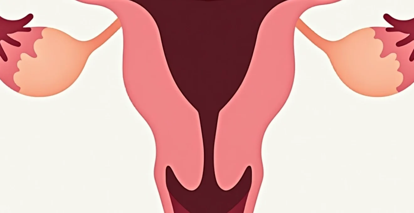
The normal anatomical position of the uterus involves a slight forward tilt, known as anteversion, where the organ rests comfortably over the bladder with the cervix pointing toward the rectum. In this configuration, the uterus forms what medical professionals often describe as a gentle curve, similar to the letter “C” when viewed from the side. This positioning allows for optimal space distribution within the pelvic cavity and typically results in minimal interference with surrounding organs during various activities, including sexual intercourse.
In contrast, a retroverted uterus demonstrates a backward tilt, with the fundus (upper portion) angled toward the spine and the cervix pointing forward toward the abdomen. This anatomical variation can be visualised as an inverted “C” shape, creating a fundamentally different spatial relationship within the pelvis. The altered positioning means that during deep penetration, the penis may directly contact the posterior wall of the uterus or compress adjacent structures, potentially triggering pain responses that wouldn’t occur with normal positioning.
Clinical classification systems for uterine retroversion degrees
Medical professionals classify uterine retroversion according to degrees of deviation from the normal anteverted position. First-degree retroversion involves a mild backward tilt that may cause minimal symptoms, whilst second and third-degree classifications represent progressively more pronounced angles. Third-degree retroversion, the most severe form, positions the uterine fundus almost horizontally against the rectum, often resulting in the most significant symptoms and complications.
This classification system proves crucial for healthcare providers when assessing treatment options and predicting symptom severity. Women with higher degrees of retroversion typically experience more pronounced sexual discomfort, increased menstrual pain, and greater likelihood of associated conditions such as endometriosis or pelvic inflammatory disease. Understanding these gradations helps both patients and clinicians develop realistic expectations for treatment outcomes and symptom management strategies.
Epidemiological data on tilted uterus occurrence in women
Recent epidemiological studies reveal that retroverted uterus affects approximately 20-30% of women globally, making it one of the most common anatomical variations of the female reproductive system. Interestingly, prevalence rates vary significantly across different populations and age groups, with some research suggesting that certain genetic factors may predispose individuals to this condition. Asian populations demonstrate slightly lower rates at around 15-20%, whilst Caucasian populations show prevalence rates closer to 25-30%.
Age-related changes also influence uterine positioning, with postmenopausal women showing increased rates of retroversion due to hormonal changes affecting ligamentous support structures. Studies indicate that approximately 40% of women over 50 develop some degree of uterine retroversion, often correlating with decreased oestrogen levels and subsequent weakening of the uterosacral ligaments that maintain normal uterine positioning.
Congenital versus acquired retroversion pathophysiology
Congenital retroversion occurs during foetal development when the uterus fails to rotate into its typical anteverted position. This developmental variation affects approximately 15% of all women and typically remains stable throughout life unless influenced by pregnancy, surgical procedures, or pathological conditions. Women born with congenital retroversion often experience consistent symptoms from menarche onwards, though many adapt to their anatomical configuration without seeking medical intervention.
Acquired retroversion develops later in life due to various pathological processes or mechanical factors. Pregnancy represents one of the most common causes, as the growing foetus can stretch uterine ligaments beyond their normal elasticity, allowing the uterus to tip backward after delivery. Additionally, conditions such as endometriosis, pelvic inflammatory disease, or large fibroids can create adhesions that pull the uterus into a retroverted position, often accompanied by more severe symptoms than congenital cases.
Physiological mechanisms linking uterine retroversion to dyspareunia
Deep penetration impact on retroverted uterine fundus
The primary mechanism by which a retroverted uterus causes sexual pain involves direct mechanical contact between the penis and the posteriorly positioned uterine fundus. During deep penetration, particularly in certain positions, the penis can compress the uterus against the rectum and surrounding pelvic structures, creating intense pressure sensations that register as sharp, cramping pain. This phenomenon, known as collision dyspareunia , represents one of the most characteristic symptoms of retroverted uterus and can significantly impact sexual satisfaction and relationship dynamics.
The intensity of this pain often correlates directly with the degree of penetration and the specific sexual positions employed. Woman-on-top positions frequently exacerbate symptoms because they allow for deeper penetration and create angles that maximise contact with the sensitive uterine fundus. Research indicates that approximately 60% of women with retroverted uterus experience significant discomfort in this position, compared to only 10% of women with normal uterine positioning.
Cervical contact and pressure distribution during intercourse
The altered cervical positioning in retroverted uterus cases creates unique pressure distribution patterns during sexual activity. Instead of the cervix pointing toward the rectum as in normal anatomy, it angles forward toward the abdomen, potentially creating increased contact with the penis during intercourse. This contact can trigger intense pain responses, particularly in women with cervical sensitivity or those who have experienced previous cervical trauma or procedures.
Additionally, the forward-angled cervix may experience increased pressure from thrusting motions, creating a secondary source of discomfort that compounds the primary uterine pain. Studies suggest that women with retroverted uterus are three times more likely to experience cervical tenderness during gynaecological examinations, indicating heightened sensitivity that extends beyond sexual activity into routine medical care.
Pelvic floor tension modifications in posterior uterine displacement
The backward positioning of a retroverted uterus significantly alters pelvic floor muscle tension and coordination patterns. Normal pelvic floor function relies on balanced muscular support that accommodates the typical anteverted uterine position. When the uterus tips backward, it can create compensatory tension in surrounding muscles, particularly the levator ani group, which may remain chronically contracted to provide additional support for the displaced organ.
This chronic muscle tension can contribute to a condition called pelvic floor dysfunction , where the muscles become unable to relax properly during sexual arousal and penetration. Women with this condition often describe a sensation of tightness or burning during intercourse, in addition to the direct uterine pain caused by their retroversion. Effective treatment often requires addressing both the anatomical positioning and the secondary muscular dysfunction through targeted physiotherapy approaches.
Ligamentous strain patterns affecting sexual comfort
The uterus is supported by several key ligamentous structures, including the uterosacral ligaments, cardinal ligaments, and pubocervical fascia. In retroverted uterus cases, these support structures experience altered tension patterns that can contribute to sexual discomfort. The uterosacral ligaments, which normally provide posterior support, may become stretched and painful when supporting a backward-tilted uterus against gravitational forces and sexual positioning.
The complex interplay between ligamentous support, muscular tension, and anatomical positioning creates a multifaceted pain syndrome that requires comprehensive assessment and individualised treatment approaches.
During sexual activity, thrusting motions can further strain these already compromised ligaments, creating referred pain patterns that extend beyond the immediate pelvic area. Many women report lower back pain, hip discomfort, and even radiating leg pain following sexual activity, symptoms that directly correlate with ligamentous strain patterns unique to retroverted uterus anatomy.
Differential diagnosis of painful intercourse in retroverted uterus cases
Endometriosis association with uterine retroversion and sexual pain
Endometriosis demonstrates a particularly strong association with uterine retroversion, with studies indicating that up to 50% of women with moderate to severe endometriosis also present with a tilted uterus. This correlation occurs because endometrial implants commonly develop on the uterosacral ligaments and posterior pelvic structures, creating adhesions that pull the uterus backward whilst simultaneously causing intense pelvic pain. The combination of retroversion and endometriosis creates a particularly challenging clinical scenario where multiple pain mechanisms overlap and compound each other.
Women with both conditions often experience cyclical pain that worsens during menstruation, deep dyspareunia that persists regardless of sexual position modifications, and chronic pelvic pain that impacts daily activities beyond sexual function. The endometrial tissue responds to hormonal fluctuations throughout the menstrual cycle, creating inflammation and pain that can make even gentle sexual contact unbearable during certain times of the month.
Pelvic inflammatory disease complications in tilted uterus patients
Pelvic inflammatory disease (PID) can both cause and complicate retroverted uterus cases through the formation of adhesions and scar tissue. When PID affects women with existing uterine retroversion, the inflammatory process often creates additional fibrous bands that further restrict uterine mobility and exacerbate sexual pain symptoms. Chronic PID may result in hydrosalpinx or tubo-ovarian complexes that physically impede normal sexual function and create persistent discomfort.
The diagnostic challenge in these cases lies in distinguishing between pain caused by the anatomical retroversion itself versus ongoing inflammatory processes. Women with PID-related retroversion typically present with additional symptoms including abnormal vaginal discharge, irregular bleeding, and fever, which help differentiate this condition from simple anatomical variation.
Adenomyosis correlation with posterior uterine position
Adenomyosis , characterised by the invasion of endometrial tissue into the uterine muscle wall, frequently coexists with uterine retroversion and can significantly amplify sexual pain symptoms. The condition causes uterine enlargement and increased sensitivity, making the mechanical contact that occurs during intercourse with a retroverted uterus even more painful and distressing. Women with adenomyosis typically experience heavy, prolonged menstrual periods alongside severe dysmenorrhea that can persist throughout the menstrual cycle.
The combination of adenomyosis and retroversion creates a particularly complex pain syndrome where hormonal, mechanical, and inflammatory factors all contribute to sexual dysfunction. Treatment approaches must address both the anatomical positioning and the underlying pathological process to achieve meaningful symptom relief and restore sexual function.
Ovarian cyst impact on retroverted uterus sexual function
Large ovarian cysts can significantly impact sexual comfort in women with retroverted uterus by occupying additional pelvic space and creating pressure points during intercourse. Functional cysts, dermoid cysts, and endometriomas all pose unique challenges when combined with posterior uterine positioning. The cysts may become compressed between the tilted uterus and penis during sexual activity, creating sharp, stabbing pains that can be severe enough to require immediate cessation of sexual activity.
Complex ovarian masses may also contribute to uterine retroversion by altering the normal anatomical relationships within the pelvis. Large cysts can physically push the uterus into a more pronounced backward tilt, worsening existing symptoms and creating new areas of tenderness and sensitivity that complicate sexual positioning and comfort strategies.
Evidence-based treatment approaches for Retroversion-Related dyspareunia
Conservative management strategies for retroversion-related sexual pain focus on symptom relief and functional improvement without surgical intervention. Pelvic floor physiotherapy represents the cornerstone of conservative treatment, utilising specialised exercises designed to improve muscular coordination and reduce compensatory tension patterns. Research demonstrates that approximately 70% of women with retroverted uterus experience significant improvement in sexual comfort following a structured 12-week pelvic floor rehabilitation programme.
Hormonal therapies may provide substantial benefit for women whose retroversion is associated with conditions like endometriosis or adenomyosis. Combined oral contraceptive pills, particularly those with anti-androgenic properties, can reduce inflammation and pain sensitivity whilst also helping to regulate menstrual cycles. GnRH agonists represent a more intensive hormonal approach reserved for severe cases where other treatments have failed to provide adequate relief.
Pessary devices offer a non-surgical mechanical solution for some women with symptomatic retroverted uterus. These silicone or plastic devices are inserted vaginally to physically support and reposition the uterus in a more forward position. However, pessaries require careful fitting and regular monitoring, as they can increase infection risk and may cause discomfort for sexual partners during intercourse.
When conservative treatments fail to provide adequate relief, surgical options become consideration. Uterine suspension procedures, including laparoscopic uterosacral ligament plication and ventral suspension techniques, can effectively reposition the uterus and provide long-term symptom relief. Success rates for these procedures approach 80-90% for appropriately selected candidates, with most women experiencing significant improvements in sexual comfort and overall quality of life.
Surgical intervention should be carefully considered only after conservative treatments have been thoroughly explored and found inadequate, as the risks and recovery requirements of surgery may outweigh the benefits for some patients.
Advanced surgical techniques now include robot-assisted procedures that offer increased precision and reduced recovery times compared to traditional open surgeries. These minimally invasive approaches typically result in shorter hospital stays, less post-operative pain, and faster return to normal sexual activity, making them increasingly attractive options for younger women seeking definitive treatment.
Sexual position modifications and biomechanical considerations
Strategic sexual positioning modifications can dramatically improve comfort and pleasure for women with retroverted uterus by reducing mechanical contact and pressure on sensitive areas. The missionary position with pillows placed under the woman’s hips often provides optimal comfort by tilting the pelvis forward and reducing direct uterine contact. This positioning allows for intimate contact whilst minimising the deep penetration that typically triggers pain in retroverted uterus cases.
Side-lying positions, particularly the spooning configuration, offer excellent alternatives that naturally limit penetration depth whilst maintaining intimacy and emotional connection. These positions allow the woman to control the angle and depth of penetration by adjusting her leg position and pelvic tilt, providing immediate feedback and adjustment capabilities that can prevent painful contact before it occurs.
- Reverse cowgirl position allows for controlled depth whilst avoiding direct fundal contact
- Modified missionary with elevated hips reduces posterior uterine pressure
- Side-entry positions provide natural depth limitation and comfort control
- Standing positions may reduce gravitational effects on retroverted anatomy
Communication between partners becomes critically important when implementing position modifications, as both individuals need to understand the anatomical challenges and work together to find mutually satisfying solutions. Many couples benefit from professional counselling or sex therapy to address the emotional and relational aspects of managing chronic sexual pain while exploring new techniques and approaches.
Biomechanical principles suggest that positions which maintain the natural lumbar curve and avoid extreme pelvic flexion typically provide the greatest comfort for women with retroverted uterus. Understanding these principles allows couples to adapt traditional positions or create entirely new approaches that prioritise comfort whilst maintaining sexual satisfaction and intimacy.
Long-term prognosis and fertility implications of uterine retroversion
The long-term outlook for women with retroverted uterus varies considerably depending on the underlying cause and associated conditions. Women with congenital retroversion who receive appropriate treatment typically experience stable symptoms that can be effectively managed through conservative approaches or surgical intervention
throughout their reproductive years and beyond. Studies tracking women with retroverted uterus over decades demonstrate that symptom severity typically remains stable when no underlying pathological conditions exist. However, hormonal changes during menopause may actually improve symptoms for some women, as decreased oestrogen levels can reduce tissue sensitivity and inflammation.
Pregnancy outcomes in women with retroverted uterus are generally excellent, with the vast majority experiencing normal conception rates and uncomplicated pregnancies. The growing uterus naturally rotates into an anteverted position during the first trimester in approximately 90% of cases, often providing temporary relief from sexual pain symptoms. This physiological repositioning occurs as the expanding uterus outgrows the pelvis and rises into the abdominal cavity, where gravitational forces naturally encourage forward positioning.
Fertility implications of uterine retroversion remain minimal in most cases, with conception rates matching those of women with normal uterine positioning. Research involving over 10,000 women attempting pregnancy showed no statistically significant difference in time to conception between those with retroverted versus anteverted uterus positioning. However, women with retroversion secondary to conditions like endometriosis or pelvic inflammatory disease may experience reduced fertility related to these underlying pathological processes rather than the uterine positioning itself.
Post-pregnancy outcomes vary depending on individual anatomical factors and the degree of ligamentous stretching that occurs during gestation. Approximately 30% of women find that their uterus returns to its original retroverted position after delivery, whilst others maintain the forward positioning achieved during pregnancy. Women who experience persistent anteversion following pregnancy often report complete resolution of their previous sexual pain symptoms, highlighting the potential for natural anatomical correction through pregnancy and childbirth.
Age-related changes in uterine positioning become increasingly relevant as women approach menopause and beyond. Declining oestrogen levels affect the integrity of supporting ligaments, potentially worsening existing retroversion or contributing to acquired cases in women who previously had normal positioning. However, these same hormonal changes may reduce pain sensitivity and inflammation, creating a complex clinical picture where anatomical worsening may paradoxically correlate with symptom improvement.
Long-term management success depends heavily on early identification of underlying conditions and implementation of appropriate treatment strategies before secondary complications develop.
Women who undergo successful surgical correction of their retroverted uterus typically maintain improved positioning for decades, with revision rates remaining below 5% when procedures are performed by experienced specialists. The durability of surgical outcomes makes these interventions particularly attractive for younger women who desire definitive symptom resolution and improved quality of life throughout their reproductive years.
Quality of life assessments conducted five years after diagnosis show that women who receive comprehensive evaluation and appropriate treatment maintain sexual satisfaction rates comparable to those without uterine positioning abnormalities. This emphasises the importance of proper medical care and the generally favourable prognosis for women who seek treatment for retroversion-related symptoms. Early intervention and partner education contribute significantly to positive long-term outcomes, highlighting the value of open communication with healthcare providers about sexual health concerns.
Regular monitoring remains important for women with retroverted uterus, particularly those with associated conditions like endometriosis or a history of pelvic inflammatory disease. Annual gynaecological examinations can identify changes in uterine positioning or the development of new pathological processes that might require treatment modification. Women who remain asymptomatic require no special monitoring beyond routine preventive care, whilst those with ongoing symptoms benefit from regular reassessment to ensure optimal management strategies.
The psychological impact of living with a retroverted uterus should not be underestimated, as chronic sexual pain can significantly affect self-esteem, relationship satisfaction, and overall mental health. Support groups and counselling services specifically addressing sexual health concerns provide valuable resources for women navigating the emotional challenges associated with this condition. Many women report that understanding the anatomical basis for their symptoms reduces anxiety and improves their ability to work with healthcare providers and partners in developing effective management strategies.