Distinguishing between peripheral neuropathy and multiple sclerosis represents one of the most challenging diagnostic dilemmas in modern neurology. Both conditions can present with overlapping symptoms including numbness, tingling, pain, and motor dysfunction, yet they fundamentally differ in their underlying pathophysiology, anatomical distribution, and treatment approaches. The complexity arises because these neurological disorders can affect similar functional domains while targeting entirely different components of the nervous system. Understanding these critical differences enables healthcare professionals to implement appropriate diagnostic strategies and therapeutic interventions, ultimately improving patient outcomes and quality of life.
Pathophysiology and disease mechanisms: peripheral neuropathy versus multiple sclerosis
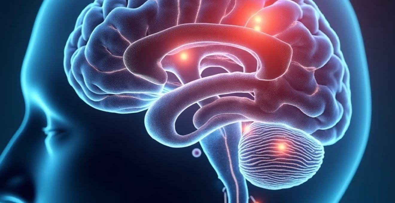
The fundamental distinction between peripheral neuropathy and multiple sclerosis lies in their anatomical targets and pathophysiological mechanisms. Peripheral neuropathy encompasses a diverse group of conditions affecting the peripheral nervous system—nerves outside the brain and spinal cord—whilst multiple sclerosis primarily targets the central nervous system, including the brain, spinal cord, and optic nerves. This anatomical differentiation serves as the cornerstone for understanding how these conditions manifest clinically and respond to various therapeutic interventions.
Multiple sclerosis involves autoimmune-mediated destruction of myelin sheaths surrounding nerve fibres within the central nervous system. The immune system mistakenly attacks oligodendrocytes, the cells responsible for producing and maintaining myelin in the CNS, leading to inflammation, demyelination, and eventual axonal damage. This process results in the characteristic plaques or lesions visible on magnetic resonance imaging, which disrupt normal neural transmission between the brain and peripheral organs.
Demyelination patterns in central versus peripheral nervous systems
The demyelination patterns in these conditions reveal fundamental differences in cellular biology and repair mechanisms. In multiple sclerosis, oligodendrocytes produce myelin for multiple axons simultaneously, meaning that damage to a single oligodendrocyte can affect numerous nerve fibres. This creates the characteristic multifocal lesion pattern observed in MS, where discrete areas of demyelination appear throughout the central nervous system.
Conversely, peripheral neuropathy involving demyelination affects Schwann cells, which maintain a one-to-one relationship with individual axons. This cellular arrangement allows for more efficient repair mechanisms in the peripheral nervous system, though the process remains dependent on the underlying cause of the neuropathy. Chronic inflammatory demyelinating polyneuropathy exemplifies this difference, often showing better recovery potential compared to progressive forms of multiple sclerosis.
Axonal degeneration in diabetic neuropathy and Charcot-Marie-Tooth disease
Axonal forms of peripheral neuropathy follow distinct pathophysiological pathways that differ markedly from the demyelinating processes in multiple sclerosis. Diabetic neuropathy , the most common form of peripheral neuropathy, results from chronic hyperglycaemia causing metabolic dysfunction, oxidative stress, and vascular compromise affecting peripheral nerves. The longest nerves are typically affected first, creating the characteristic distal sensorimotor pattern.
Hereditary neuropathies like Charcot-Marie-Tooth disease demonstrate genetic mutations affecting various aspects of peripheral nerve function, including myelin production, axonal transport, and cellular metabolism. These conditions highlight how peripheral neuropathy can result from intrinsic cellular dysfunction rather than external inflammatory processes, contrasting sharply with the autoimmune mechanisms underlying multiple sclerosis.
Autoimmune inflammatory cascades in Guillain-Barré syndrome
Guillain-Barré syndrome represents an acute autoimmune peripheral neuropathy that provides insight into how immune-mediated nerve damage differs between peripheral and central nervous systems. The condition typically follows infections and involves molecular mimicry, where antibodies against infectious agents cross-react with peripheral nerve components. This autoimmune response targets various peripheral nerve antigens, including gangliosides and myelin proteins.
The inflammatory cascade in Guillain-Barré syndrome demonstrates how peripheral nerves respond to immune attack differently than central nervous system tissues. Peripheral nerves maintain greater regenerative capacity and can recover more completely, partly due to the presence of Schwann cells and the absence of inhibitory factors present in the central nervous system environment.
Oligodendrocyte destruction in Relapsing-Remitting MS
The pathophysiology of relapsing-remitting multiple sclerosis involves complex interactions between activated T-cells, B-cells, and innate immune cells that breach the blood-brain barrier. Oligodendrocyte destruction occurs through both direct cellular cytotoxicity and inflammatory mediator release, including cytokines, chemokines, and complement proteins. This process creates acute inflammatory lesions that can partially remyelinate during remission phases.
The central nervous system’s limited regenerative capacity compared to peripheral nerves explains why multiple sclerosis often leads to progressive disability over time. Oligodendrocyte precursor cells attempt to repair damaged myelin, but this process becomes increasingly inefficient with repeated inflammatory attacks, leading to chronic demyelination and axonal loss.
Clinical presentation and symptom differentiation patterns
The clinical presentations of peripheral neuropathy and multiple sclerosis exhibit distinct patterns that reflect their underlying anatomical distributions and pathophysiological mechanisms. Recognition of these patterns enables healthcare professionals to develop appropriate differential diagnostic strategies and implement targeted therapeutic approaches. Understanding symptom evolution, anatomical distribution, and temporal characteristics provides crucial insights for distinguishing between these neurological conditions.
Distal sensorimotor deficits in Length-Dependent polyneuropathy
Length-dependent polyneuropathy demonstrates the characteristic distal-to-proximal progression that typifies many peripheral neuropathies. Symptoms typically begin in the toes and fingertips, gradually ascending proximally as longer nerve fibres become progressively affected. This pattern reflects the vulnerability of longer axons to metabolic disturbances, toxic exposures, and nutritional deficiencies.
The stocking-glove distribution of sensory loss in peripheral neuropathy contrasts markedly with the often asymmetric and multifocal symptom patterns observed in multiple sclerosis. Patients with diabetic polyneuropathy commonly experience burning pain, numbness, and tingling that begins in the feet and may progress to involve the hands as the condition advances. Motor symptoms typically appear later and follow a similar distal predominance.
Trigeminal neuralgia and lhermitte’s sign in multiple sclerosis
Multiple sclerosis can produce distinctive neurological signs that rarely occur in peripheral neuropathy. Trigeminal neuralgia in MS patients results from demyelinating lesions affecting the trigeminal nerve’s central pathways within the brainstem. This condition produces characteristic sharp, electric shock-like pain episodes triggered by light touch or movement of facial muscles.
Lhermitte’s sign represents another pathognomonic feature of multiple sclerosis, consisting of electric shock-like sensations radiating down the spine and extremities triggered by neck flexion. This symptom results from demyelinated cervical spinal cord lesions that become mechanically sensitive to movement. Such central nervous system signs help differentiate MS from peripheral neuropathies, which typically lack these characteristic features.
Stocking-glove distribution versus disseminated CNS lesions
The anatomical distribution of symptoms provides one of the most reliable methods for distinguishing peripheral neuropathy from multiple sclerosis. Peripheral neuropathy commonly produces symmetric symptoms following specific nerve distributions, such as the stocking-glove pattern in distal sensorimotor polyneuropathy or dermatomal distributions in radiculopathies.
Multiple sclerosis lesions create symptom patterns that reflect their random distribution throughout the central nervous system. Patients may experience unilateral sensory loss, monocular vision changes, or asymmetric motor weakness that doesn’t conform to peripheral nerve territories. The temporal evolution also differs, with MS often showing relapsing-remitting patterns compared to the typically progressive nature of most peripheral neuropathies.
Uhthoff’s phenomenon and Temperature-Related MS exacerbations
Uhthoff’s phenomenon represents a unique characteristic of multiple sclerosis involving temporary worsening of neurological symptoms with increased body temperature. This temperature sensitivity results from impaired conduction in demyelinated central nervous system fibres, where even slight temperature increases can block action potential propagation. Heat exposure, fever, exercise, or hot baths can trigger symptom exacerbations that typically resolve with cooling.
Peripheral neuropathies rarely demonstrate similar temperature sensitivity patterns. While some patients with peripheral neuropathy may experience weather-related symptom fluctuations, these typically involve pain sensitivity to cold temperatures rather than the heat-induced conduction blocks characteristic of multiple sclerosis. This distinction can provide valuable diagnostic information when evaluating patients with overlapping neurological symptoms.
Carpal tunnel syndrome and focal entrapment neuropathies
Focal entrapment neuropathies like carpal tunnel syndrome demonstrate how peripheral nerve dysfunction can create localised symptoms that differ significantly from the disseminated lesions of multiple sclerosis. Carpal tunnel syndrome produces characteristic median nerve distribution symptoms, including nocturnal hand pain, thumb and finger numbness, and eventually thenar muscle weakness.
These focal neuropathies result from mechanical compression of peripheral nerves at anatomically vulnerable sites. The symptoms remain confined to specific nerve territories and often respond to local treatments such as splinting or surgical decompression. This contrasts with multiple sclerosis symptoms, which reflect central nervous system lesion locations and require systemic immunomodulatory therapies.
Advanced neuroimaging and electrodiagnostic testing protocols
Contemporary diagnostic approaches for differentiating peripheral neuropathy from multiple sclerosis rely heavily on advanced neuroimaging techniques and electrodiagnostic studies. These sophisticated testing modalities provide objective evidence of nervous system dysfunction and help localise pathology to specific anatomical compartments. The integration of multiple diagnostic technologies enables clinicians to establish definitive diagnoses and monitor disease progression with unprecedented precision.
Gadolinium-enhanced MRI detection of Blood-Brain barrier disruption
Gadolinium-enhanced magnetic resonance imaging serves as the gold standard for detecting active inflammation in multiple sclerosis. The contrast agent reveals areas where the blood-brain barrier has been compromised by inflammatory processes, appearing as enhancing lesions on T1-weighted sequences. These enhancing lesions indicate acute inflammatory activity and help distinguish active from chronic lesions in MS patients.
The presence of gadolinium enhancement provides crucial information for treatment decisions and prognosis in multiple sclerosis. Active enhancing lesions suggest ongoing inflammatory activity that may respond to immunomodulatory therapies, whilst the absence of enhancement in chronic lesions indicates established tissue damage with limited therapeutic potential. This imaging capability remains largely irrelevant in peripheral neuropathy diagnosis, where blood-nerve barrier disruption occurs through different mechanisms.
Nerve conduction velocity studies and F-Wave analysis
Electrodiagnostic testing provides the primary objective assessment tool for peripheral neuropathy evaluation. Nerve conduction studies measure the speed and amplitude of electrical signal transmission through peripheral nerves, revealing both demyelinating and axonal pathology patterns. Demyelinating neuropathies typically show slowed conduction velocities with preserved amplitudes, whilst axonal neuropathies demonstrate reduced amplitudes with relatively preserved conduction speeds.
F-wave studies assess the integrity of the entire peripheral nerve pathway, including proximal segments that may be inaccessible to routine conduction studies. Prolonged F-wave latencies or absent responses can indicate proximal nerve involvement, helping differentiate between peripheral neuropathy and central nervous system pathology. These studies remain normal in pure multiple sclerosis cases without concurrent peripheral nerve involvement.
Nerve conduction studies provide objective, quantitative assessment of peripheral nerve function and serve as the cornerstone for differentiating peripheral neuropathy from central nervous system disorders.
T2-hyperintense lesions in periventricular white matter
T2-weighted magnetic resonance imaging reveals characteristic lesion patterns that help distinguish multiple sclerosis from other neurological conditions. Periventricular white matter lesions in MS typically demonstrate specific morphological features, including ovoid shape, perpendicular orientation to ventricular surfaces, and preferential involvement of certain anatomical regions such as the corpus callosum and brainstem.
The distribution and characteristics of T2 hyperintense lesions follow established diagnostic criteria for multiple sclerosis, including the McDonald criteria. These lesions must demonstrate dissemination in space and time to support an MS diagnosis. Peripheral neuropathy patients typically show normal central nervous system imaging, though some may have incidental age-related white matter changes that must be distinguished from pathological lesions.
Electromyography patterns in axonal versus demyelinating neuropathy
Electromyographic assessment reveals distinctive patterns that help characterise the underlying pathophysiology in peripheral neuropathy. Axonal neuropathies demonstrate reduced recruitment of motor units with normal or mildly prolonged motor unit potentials, reflecting loss of functioning nerve fibres. Chronic axonal damage may show large, long-duration, polyphasic motor unit potentials indicating reinnervation attempts.
Demyelinating neuropathies produce different electromyographic patterns, often showing normal motor unit morphology with reduced recruitment due to conduction blocks rather than axonal loss. Temporal dispersion and conduction blocks become evident on nerve conduction studies, helping differentiate demyelinating from axonal pathology. These electrodiagnostic patterns remain absent in pure multiple sclerosis cases.
Cerebrospinal fluid analysis and laboratory biomarkers
Cerebrospinal fluid analysis provides valuable diagnostic information for distinguishing multiple sclerosis from peripheral neuropathy, particularly in cases where clinical presentation and imaging findings remain ambiguous. The CSF reflects pathological processes occurring within the central nervous system and can reveal inflammatory markers, antibodies, and cellular changes specific to different neurological conditions. Lumbar puncture remains an important diagnostic tool when combined with appropriate clinical and radiological assessments.
Multiple sclerosis demonstrates characteristic CSF abnormalities that support the diagnosis and provide prognostic information. Oligoclonal bands represent immunoglobulin G antibodies produced intrathecally within the central nervous system, appearing in approximately 90-95% of MS patients. These bands indicate ongoing immune activation within the CNS and remain absent in most peripheral neuropathy cases, providing crucial differential diagnostic information.
The CSF protein concentration and cellular composition also differ between these conditions. Multiple sclerosis typically shows normal or mildly elevated protein levels (usually less than 100 mg/dL) with modest lymphocytic pleocytosis. Peripheral neuropathy may produce different CSF patterns depending on the underlying aetiology, with some inflammatory neuropathies causing marked protein elevation due to blood-nerve barrier dysfunction. Guillain-Barré syndrome characteristically produces cytoalbuminous dissociation with markedly elevated protein levels but normal cell counts.
Advanced CSF biomarkers continue to evolve, providing enhanced diagnostic capabilities for distinguishing between neurological conditions. Neurofilament light chain levels reflect axonal damage and can be elevated in both conditions, though the patterns and implications differ. CSF neurofilament levels in MS correlate with disease activity and progression, whilst in peripheral neuropathy, they may indicate the severity of axonal involvement. IgG index and synthesis rates provide additional measures of intrathecal antibody production specific to multiple sclerosis.
Cerebrospinal fluid oligoclonal bands provide highly specific evidence of central nervous system inflammation and remain one of the most reliable laboratory markers for distinguishing multiple sclerosis from peripheral neuropathy.
Pharmacological treatment approaches and Disease-Modifying therapies
The therapeutic landscape for peripheral neuropathy and multiple sclerosis reflects their fundamentally different pathophysiological mechanisms and treatment goals. Disease-modifying therapies for multiple sclerosis focus primarily on immunomodulation and immunosuppression to reduce inflammatory activity within the central nervous system. These treatments include interferon beta preparations, glatiramer acetate, dimethyl fumarate, and various monoclonal antibodies targeting specific immune system components.
Peripheral neuropathy management emphasises addressing underlying causes when possible and providing symptomatic relief for neuropathic pain and functional impairment. Treatment approaches vary significantly depending on the specific type of neuropathy, ranging from glucose control in diabetic neuropathy to vitamin supplementation in nutritional deficiencies. Immunosuppressive treatments may be appropriate for inflammatory neurop
athies such as chronic inflammatory demyelinating polyneuropathy and Guillain-Barré syndrome.
The symptomatic management of peripheral neuropathy emphasises pain control using medications specifically designed for neuropathic pain. These include anticonvulsants like gabapentin and pregabalin, tricyclic antidepressants such as amitriptyline, and newer agents like duloxetine. Unlike multiple sclerosis treatments that target disease progression, peripheral neuropathy medications primarily focus on improving quality of life and functional capacity through symptom relief.
Multiple sclerosis disease-modifying therapies demonstrate remarkable complexity and sophistication, reflecting our evolving understanding of immune system dysfunction in this condition. Newer treatments like natalizumab, alemtuzumab, and ocrelizumab target specific immune cell populations or adhesion molecules, providing more precise therapeutic interventions. These treatments require careful monitoring for potential complications, including progressive multifocal leukoencephalopathy and infusion reactions.
The treatment paradigms also differ significantly in their monitoring requirements and long-term management strategies. Multiple sclerosis patients require regular MRI surveillance to assess treatment efficacy and disease progression, whilst peripheral neuropathy management typically relies on clinical assessment and functional outcome measures. Treatment duration varies considerably, with MS therapies often requiring indefinite continuation compared to peripheral neuropathy treatments that may be adjusted based on symptom response and underlying cause resolution.
Disease-modifying therapies for multiple sclerosis have revolutionised patient outcomes by reducing relapse rates and slowing disability progression, whilst peripheral neuropathy treatments focus primarily on symptom management and addressing reversible underlying causes.
Prognosis and long-term neurological outcomes assessment
The long-term prognosis for patients with peripheral neuropathy versus multiple sclerosis varies dramatically based on underlying aetiology, treatment response, and individual patient factors. Multiple sclerosis prognosis has improved significantly with the advent of disease-modifying therapies, though the condition remains progressive in many patients. Relapsing-remitting MS may transition to secondary progressive forms, whilst primary progressive MS typically shows steady deterioration from onset.
Peripheral neuropathy outcomes depend heavily on the underlying cause and timing of intervention. Diabetic neuropathy may stabilise or improve with optimal glucose control, whilst hereditary neuropathies typically progress slowly over decades. Inflammatory neuropathies often show excellent recovery potential, particularly when treatment begins early in the disease course. This variability in prognosis reflects the diverse pathophysiological mechanisms underlying different peripheral neuropathy subtypes.
Functional outcome assessments require different approaches for these conditions. Multiple sclerosis disability is commonly measured using the Expanded Disability Status Scale (EDSS), which emphasises ambulatory function and reflects the condition’s impact on mobility and daily activities. Peripheral neuropathy assessment focuses more on sensory function, pain levels, and specific functional deficits related to the affected nerve distributions.
The quality of life implications differ significantly between these conditions. Multiple sclerosis patients often experience cognitive changes, fatigue, and mood disorders that extend beyond physical disability. Peripheral neuropathy typically produces more localised symptoms, though chronic pain and sensory loss can significantly impact daily functioning and psychological well-being. Understanding these different prognostic patterns enables healthcare providers to counsel patients appropriately and establish realistic treatment goals.
Rehabilitation strategies also vary based on the specific neurological deficits present in each condition. Multiple sclerosis rehabilitation emphasises maintaining function during relapses, managing fatigue, and adapting to progressive disability. Peripheral neuropathy rehabilitation focuses on fall prevention, pain management, and compensatory strategies for sensory loss. Both conditions benefit from multidisciplinary approaches, though the specific interventions and therapeutic goals differ substantially based on the underlying pathophysiology and expected disease course.