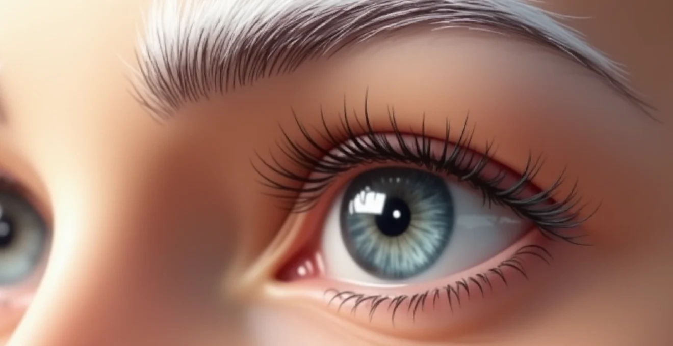
Discovering a solitary white hair sprouting from your eyebrow can trigger a cascade of questions about health, genetics, and ageing. This seemingly minor cosmetic change often represents a complex interplay of biological processes occurring within individual hair follicles. Unlike the gradual greying pattern typically observed across the scalp, isolated white eyebrow hairs present a unique phenomenon that warrants deeper understanding. The appearance of poliosis —the medical term for localised hair depigmentation—in a single eyebrow hair can indicate various underlying mechanisms, from benign age-related changes to more significant systemic conditions.
Melanin production disruption and follicular pigmentation loss
The transformation of a pigmented hair to white fundamentally stems from disrupted melanin synthesis within the hair follicle’s matrix. Melanocytes, specialised cells responsible for producing melanin pigments, can experience functional impairment through multiple pathways. When these cellular factories cease operation or operate inefficiently, the resulting hair shaft emerges devoid of its characteristic colouration. This process differs significantly from complete hair loss, as the follicle remains structurally intact while losing only its pigment-producing capacity.
Understanding melanin production requires examining the intricate biochemical cascade that normally occurs during hair growth cycles. The anagen phase, representing active hair growth, depends heavily on melanocyte activity coordinated with keratinocyte proliferation. Disruption at any stage of this carefully orchestrated process can result in depigmented hair emergence , creating the stark white appearance that catches attention in otherwise uniformly coloured eyebrows.
Tyrosinase enzyme deficiency in individual hair follicles
Tyrosinase represents the rate-limiting enzyme in melanin biosynthesis, converting tyrosine amino acids into melanin precursors. Individual follicles may experience localised tyrosinase deficiency due to genetic mutations, oxidative damage, or acquired enzymatic dysfunction. This deficiency doesn’t necessarily affect neighbouring follicles, explaining why single white hairs can appear amidst otherwise normally pigmented areas. The enzyme’s copper-dependent nature makes it particularly susceptible to trace element imbalances that might affect specific follicular units while sparing others.
Oxidative stress impact on melanocyte function
Reactive oxygen species accumulation within hair follicles creates a hostile environment for melanocyte survival and function. These cellular stressors, generated through normal metabolic processes or external factors, can overwhelm the follicle’s antioxidant defence mechanisms. When oxidative stress exceeds the protective capacity of catalase, superoxide dismutase, and other defensive enzymes, melanocytes may undergo apoptosis or functional senescence. This process can affect individual follicles unpredictably, contributing to the sporadic appearance of isolated white hairs.
Age-related melanin synthesis decline in eyebrow follicles
Chronological ageing brings inevitable changes to follicular stem cell populations and their regenerative capacity. The melanocyte stem cell niche, located in the bulge region of hair follicles, experiences gradual depletion over time. As these stem cells become exhausted or dysfunctional, their ability to replenish active melanocytes during each hair cycle diminishes. Eyebrow follicles, despite their typically slower cycling compared to scalp hair, remain vulnerable to this age-related decline, though the timing varies considerably between individuals and even between follicles on the same person.
Environmental UV damage to follicular melanin cells
Ultraviolet radiation exposure affects eyebrow follicles differently than scalp hair due to their exposed position and lack of protective coverage. UV damage accumulates over years, creating DNA lesions and protein modifications that compromise melanocyte viability. The eyebrow region’s prominence makes it particularly susceptible to direct solar exposure, potentially accelerating follicular ageing and pigment loss. This environmental factor can create a patchwork effect where some follicles experience greater cumulative damage than others, leading to irregular depigmentation patterns.
Poliosis localised: clinical manifestation in single eyebrow hairs
Poliosis encompasses various conditions characterised by localised hair depigmentation, ranging from congenital syndromes to acquired disorders. When manifesting as isolated eyebrow hair whitening, poliosis often represents the earliest visible sign of underlying pathological processes. The clinical significance of single white eyebrow hairs extends beyond cosmetic concerns, potentially indicating systemic conditions requiring medical evaluation. Dermatologists increasingly recognise isolated poliosis as a valuable diagnostic clue, particularly when accompanied by other subtle clinical findings.
The differential diagnosis of localised poliosis involves careful consideration of patient history, family background, and associated symptoms. What might initially appear as a benign age-related change could represent the prodromal phase of autoimmune conditions, genetic disorders, or inflammatory processes. Early recognition and appropriate investigation of isolated eyebrow depigmentation can facilitate timely intervention when underlying treatable conditions exist.
Dermatological classification of isolated white hair syndrome
Medical literature recognises isolated white hair syndrome as a distinct clinical entity characterised by sporadic, non-progressive hair depigmentation without associated systemic features. This classification helps distinguish benign cases from those requiring further investigation. Diagnostic criteria typically include the absence of skin depigmentation, normal hair shaft structure under microscopic examination, and lack of inflammatory changes in surrounding tissue. The syndrome’s benign nature provides reassurance for patients experiencing isolated eyebrow hair whitening without other concerning features.
Vogt-koyanagi-harada disease early indicators
Vogt-Koyanagi-Harada disease, a multisystem inflammatory condition, can initially manifest through subtle changes including isolated hair depigmentation. Early-stage disease may present with single white eyebrow hairs before developing the characteristic uveal inflammation, neurological symptoms, or cutaneous changes. Recognition of this possibility emphasises the importance of comprehensive evaluation when patients report recent onset of localised poliosis, particularly if accompanied by visual disturbances, hearing changes, or headaches. Ophthalmological examination becomes crucial in suspected cases to detect subclinical ocular involvement.
Waardenburg syndrome type 1 segmental presentation
Waardenburg syndrome, a genetic condition affecting neural crest cell development, occasionally presents with segmental features including isolated eyebrow depigmentation. Type 1 Waardenburg syndrome may manifest subtly through single white hairs before more obvious features like heterochromia or hearing impairment become apparent. Genetic counselling becomes relevant when family history suggests hereditary patterns of pigmentary anomalies, sensory deficits, or craniofacial variations. Molecular testing for PAX3 mutations can confirm suspected cases and inform reproductive planning.
Vitiligo follicular pattern recognition
Vitiligo’s follicular variant can initially affect hair pigmentation before obvious skin depigmentation develops. Single white eyebrow hairs may represent the earliest manifestation of this autoimmune condition, preceding visible skin changes by months or years. Dermatoscopic examination of affected areas may reveal subtle perifollicular hypopigmentation invisible to naked eye inspection. Understanding this progression pattern helps clinicians recognise early vitiligo and implement appropriate monitoring protocols for disease progression.
Nutritional deficiencies affecting melanogenesis pathways
Nutritional status profoundly influences melanin production through multiple biochemical pathways. Deficiencies in specific vitamins, minerals, and amino acids can disrupt normal pigmentation processes, potentially causing isolated hair depigmentation. The complex nutritional requirements for melanogenesis mean that subtle deficiencies might affect individual follicles variably, explaining why some hairs lose pigmentation while others remain unaffected. Modern dietary patterns, absorption disorders, and metabolic conditions can all contribute to nutritional inadequacies affecting hair pigmentation.
Copper deficiency represents one of the most significant nutritional causes of hair depigmentation, as this trace element serves as an essential cofactor for tyrosinase enzyme function. Iron deficiency, while more commonly associated with hair loss, can also impair melanin synthesis through its role in enzymatic processes. B-vitamin complexes, particularly B12, folate, and biotin, contribute to normal melanocyte function and hair pigmentation. Comprehensive nutritional assessment becomes particularly important when evaluating patients with recent onset of hair depigmentation, especially if accompanied by other signs of nutrient deficiency.
Nutritional interventions targeting melanogenesis pathways show promise in preventing further hair depigmentation, though reversal of existing white hairs remains challenging once melanocyte populations are significantly depleted.
Autoimmune conditions targeting hair follicle melanocytes
Autoimmune mechanisms can selectively target melanocytes within hair follicles, sparing surrounding structures while eliminating pigment-producing cells. These conditions operate through various immunological pathways, including cellular immunity, humoral responses, and inflammatory cascades. The selective nature of autoimmune targeting explains why individual follicles may be affected while neighbouring structures remain normal. Understanding these mechanisms provides insight into treatment possibilities and prognosis for patients experiencing autoimmune-mediated hair depigmentation.
The relationship between systemic autoimmune conditions and localised hair depigmentation remains an active area of research. Patients with established autoimmune disorders show increased incidence of poliosis compared to the general population, suggesting shared pathophysiological mechanisms. Cross-reactive antibodies may target antigens expressed both in melanocytes and other tissues affected by systemic autoimmune conditions. This overlap emphasises the importance of screening for systemic autoimmune markers when evaluating patients with unexplained hair depigmentation.
Alopecia areata selective pigment cell destruction
Alopecia areata’s inflammatory process can selectively target melanocytes while sparing hair follicle stem cells, resulting in regrowth of depigmented hair. This phenomenon occurs when the autoimmune attack affects melanocyte populations more severely than keratinocyte stem cells responsible for hair shaft production. Patients may notice white or grey regrowth in areas previously affected by alopecia areata, reflecting the differential vulnerability of follicular cell populations. Understanding this selective targeting helps predict recovery patterns and set appropriate patient expectations regarding cosmetic outcomes.
Thyroid dysfunction impact on eyebrow pigmentation
Thyroid hormones regulate multiple aspects of hair follicle function, including melanocyte activity and pigment production. Both hyperthyroidism and hypothyroidism can disrupt normal melanogenesis, though through different mechanisms. Thyroid dysfunction affects the hair growth cycle timing, potentially creating asynchrony between keratinocyte proliferation and melanocyte activity. This temporal mismatch can result in depigmented hair production during specific growth phases. Eyebrow hair loss represents a classical sign of thyroid dysfunction, but isolated pigmentation changes may precede obvious structural changes by months.
Systemic lupus erythematosus follicular manifestations
Systemic lupus erythematosus can affect hair follicles through multiple pathways, including direct immune complex deposition, complement activation, and inflammatory mediator release. Follicular manifestations may include structural damage, cicatricial alopecia, or isolated pigment loss. Lupus-associated poliosis often occurs in conjunction with other cutaneous lupus features, but isolated hair depigmentation can represent an early or subtle manifestation. Antinuclear antibody testing and specific lupus markers help identify patients requiring systemic evaluation and treatment.
Genetic polymorphisms in melanin regulatory genes
Genetic variations in melanin regulatory pathways contribute significantly to individual differences in hair pigmentation patterns and timing of age-related changes. Polymorphisms in genes such as MC1R, TYRP1, and KITLG influence melanocyte function, pigment production efficiency, and response to environmental stressors. These genetic factors help explain why some individuals develop isolated white hairs at young ages while others maintain uniform pigmentation well into advanced years. Understanding genetic contributions provides valuable insights for predicting pigmentation patterns and developing targeted interventions.
The interaction between genetic susceptibility and environmental factors creates complex patterns of hair depigmentation that vary significantly between individuals. Epigenetic modifications can alter gene expression patterns in response to stress, nutrition, or exposure factors, potentially affecting melanocyte function in specific follicles. Pharmacogenomic considerations become relevant when evaluating treatment options, as genetic variations influence responses to interventions targeting melanogenesis pathways. Personalised approaches based on genetic profiling may represent the future direction for managing hair pigmentation disorders.
Recent genome-wide association studies have identified over 180 genetic loci associated with hair pigmentation, demonstrating the complex polygenic nature of melanin regulation and individual variation in pigmentation patterns.
Pharmaceutical-induced depigmentation of eyebrow hair
Numerous medications can affect hair pigmentation through various mechanisms, including direct melanotoxicity, interference with enzymatic pathways, or disruption of cellular metabolism. Chemotherapy agents represent the most obvious example, but many common medications can subtly affect pigment production. Antimalarial drugs, certain antibiotics, and psychotropic medications have all been associated with hair depigmentation. The timing and pattern of medication-induced changes vary considerably, with some effects appearing during treatment while others manifest months after discontinuation.
Drug-induced hair depigmentation often demonstrates unique characteristics that help distinguish it from other causes. The temporal relationship between medication initiation and pigment changes provides important diagnostic clues, though delayed effects can complicate this assessment. Dose-dependent relationships may exist for certain medications, with higher doses or longer treatment durations increasing depigmentation risk. Fortunately, medication-induced changes are often reversible upon drug discontinuation, though recovery may require months or years depending on the specific agent and duration of exposure. Healthcare providers should consider medication history when evaluating patients with unexplained hair depigmentation, particularly when other obvious causes are absent.
Comprehensive medication review, including over-the-counter supplements and topical preparations, remains essential when investigating unexplained hair depigmentation, as even seemingly innocuous products can affect melanocyte function through systemic or local mechanisms.