Wavy thumbnails represent more than just a cosmetic concern—they serve as valuable diagnostic indicators of underlying health conditions and nutritional imbalances. When the normally smooth nail plate develops ridges, dents, or undulating surfaces, it signals disruptions in the nail matrix’s cellular production process. These changes can manifest as longitudinal ridges running from cuticle to tip, or as horizontal depressions known as Beau’s lines. Understanding the complex interplay between systemic health, environmental factors, and nail morphology enables healthcare professionals and individuals to identify potential issues before they become more serious complications.
The nail matrix, located beneath the cuticle, functions as the nail’s growth centre, continuously producing keratin-rich cells that form the visible nail plate. When this delicate system encounters interference—whether from nutritional deficiencies, autoimmune conditions, trauma, or metabolic disorders—the resulting nail deformities can persist for months as the affected areas grow outward. Recognising these patterns and their underlying causes represents the first step toward effective treatment and prevention strategies.
Understanding nail plate dystrophy: wavy ridges and beau’s lines formation
Longitudinal ridging patterns and nail matrix dysfunction
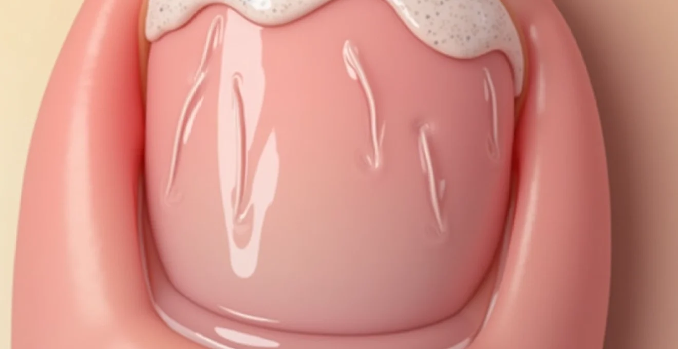
Longitudinal ridges appear as vertical lines extending from the nail base toward the free edge, creating a washboard-like texture across the thumbnail surface. These ridges typically develop when the nail matrix experiences uneven cell production, often related to age-related changes in keratinocyte synthesis. The severity of longitudinal ridging correlates directly with the extent of matrix dysfunction, ranging from barely perceptible lines to pronounced grooves that significantly alter nail appearance.
Matrix dysfunction occurs through several mechanisms, including reduced blood flow to the nail bed, decreased cellular regeneration rates, and alterations in protein synthesis pathways. Environmental factors such as repetitive trauma, chemical exposure, or mechanical stress can exacerbate these changes, leading to more pronounced ridging patterns. The thumb, being the most frequently used digit, often shows the earliest and most severe manifestations of these dystrophic changes.
Transverse wave deformities and growth interruption cycles
Transverse ridges, commonly referred to as Beau’s lines, manifest as horizontal depressions across the nail plate, indicating temporary interruptions in nail growth. These formations occur when the nail matrix temporarily ceases or significantly reduces cell production, often in response to acute illness, severe stress, or nutritional crises. The depth and width of these lines correspond to the duration and severity of the growth interruption.
The formation process follows a predictable timeline: growth interruption occurs at the matrix level, creating a temporary reduction in nail thickness. As normal growth resumes, the affected area moves distally with the growing nail plate, becoming visible approximately 2-3 months after the initial triggering event. Multiple Beau’s lines suggest recurring episodes of growth interruption, warranting investigation into chronic underlying conditions.
Onychorrhexis versus normal Age-Related nail changes
Onychorrhexis represents a specific type of nail dystrophy characterised by longitudinal splitting and ridging, distinct from normal age-related changes in nail structure. While ageing naturally produces some degree of vertical ridging due to decreased keratin production and reduced nail plate flexibility, onychorrhexis involves actual structural fragmentation of the nail layers. This condition often affects the thumbnail preferentially due to its exposure to repeated mechanical stress.
Distinguishing between pathological onychorrhexis and normal ageing requires careful examination of ridge depth, associated symptoms, and patient history. Normal age-related ridging typically presents as shallow, uniform lines without accompanying nail splitting or brittleness. Pathological ridging, however, demonstrates irregular depths, potential nail plate separation, and often correlates with systemic health issues or prolonged environmental exposures.
Koilonychia development and iron deficiency correlations
Koilonychia, characterised by concave or spoon-shaped nails, represents an advanced manifestation of chronic iron deficiency affecting nail structure. While not directly causing wavy patterns, koilonychia often co-exists with ridging abnormalities, creating complex nail deformities. The thumbnail’s size and prominence make koilonychia particularly noticeable in this digit, often serving as an early indicator of systemic iron metabolism disorders.
The development of koilonychia involves progressive thinning of the nail plate combined with altered keratin cross-linking patterns. Iron deficiency impairs cellular oxygen transport to the nail matrix, reducing the efficiency of keratinocyte production and compromising nail structural integrity. Early recognition of koilonychia alongside ridging patterns can facilitate prompt diagnosis and treatment of underlying iron deficiency states before more serious complications develop.
Systemic medical conditions triggering nail surface irregularities
Psoriatic arthritis and nail psoriasis manifestations
Psoriatic arthritis frequently produces distinctive nail changes, including pitting, ridging, and onycholysis, with thumb involvement occurring in approximately 80% of affected patients. The inflammatory processes characteristic of psoriasis directly impact the nail matrix and nail bed, creating irregular keratinocyte production patterns that manifest as surface irregularities. These changes often precede joint symptoms by months or years, making nail examination crucial for early diagnosis.
Nail psoriasis presents with multiple morphological changes simultaneously: punctate pitting creates small depressions across the nail surface, while longitudinal ridging develops from chronic inflammation affecting matrix function. The combination of these features with salmon-coloured patches beneath the nail plate creates pathognomonic signs of psoriatic involvement. Severe cases may demonstrate complete nail dystrophy with crumbling and loss of nail plate integrity.
Thyroid dysfunction effects on keratinocyte production
Thyroid disorders significantly impact nail growth rates and structural integrity through their effects on cellular metabolism and protein synthesis. Hypothyroidism typically produces slow-growing, thick nails with prominent longitudinal ridging, while hyperthyroidism often results in thin, fragile nails with increased growth rates but poor structural quality. The thumbnail’s size makes these changes particularly apparent to both patients and clinicians.
The mechanism involves thyroid hormone regulation of keratinocyte proliferation and differentiation within the nail matrix. Disrupted hormone levels alter the normal cellular turnover patterns, creating uneven nail plate formation and surface irregularities. Thyroid-related nail changes often improve gradually following appropriate hormone replacement therapy or antithyroid treatment, though complete resolution may require 6-12 months of nail regrowth.
Autoimmune disorders: lichen planus and alopecia areata
Lichen planus affecting the nails creates distinctive ridging patterns through inflammatory destruction of the nail matrix. This autoimmune condition produces longitudinal ridging, splitting, and potential permanent nail loss if left untreated. The inflammatory infiltrate specifically targets the matrix region, creating localised areas of reduced keratinocyte production that manifest as surface irregularities.
Alopecia areata, while primarily affecting hair follicles, commonly produces nail changes including fine pitting and ridging patterns. Approximately 20% of patients with alopecia areata develop nail abnormalities, with thumbnails showing changes in 60% of affected individuals. The shared embryological origin of hair follicles and nail structures explains this association, as autoimmune processes targeting keratinocyte stem cells affect both systems simultaneously.
Cardiovascular disease indicators through nail morphology
Cardiovascular conditions often manifest through nail changes due to altered peripheral circulation and oxygen delivery to the nail matrix. Chronic heart failure, peripheral arterial disease, and other circulatory disorders can produce characteristic ridging patterns alongside other nail abnormalities such as clubbing or cyanosis. The thumbnail’s vascular supply makes it particularly sensitive to circulatory changes.
The pathophysiology involves reduced oxygen and nutrient delivery to the rapidly dividing cells of the nail matrix, compromising normal keratinocyte production and organisation. Early cardiovascular disease may present with subtle nail changes before more obvious cardiac symptoms develop, making nail examination a valuable screening tool. Splinter haemorrhages beneath the nail plate may accompany ridging in cases of endocarditis or other inflammatory cardiovascular conditions.
Nutritional deficiencies and metabolic factors in nail deformation
Biotin deficiency and keratin synthesis disruption
Biotin serves as an essential cofactor for enzymes involved in keratin synthesis, making its deficiency particularly relevant to nail health. Biotin deficiency manifests as brittle, ridged nails with increased fragility and splitting tendencies. The thumbnail, being subject to frequent mechanical stress, often shows the most pronounced changes in biotin-deficient states. Clinical studies demonstrate improvement in nail quality within 6-8 weeks of biotin supplementation at therapeutic doses of 2.5-10 mg daily.
The enzymatic pathways affected by biotin deficiency include acetyl-CoA carboxylase and pyruvate carboxylase, both crucial for cellular energy production and protein synthesis. When these pathways function suboptimally, keratinocyte production decreases and structural protein quality diminishes. Supplementation protocols must consider individual absorption rates and concurrent nutritional status, as other B-complex vitamins often require simultaneous replacement.
Zinc malabsorption and protein metabolism disorders
Zinc deficiency creates distinctive nail abnormalities including transverse white bands, ridging, and delayed growth rates. The mineral’s role in protein synthesis and cellular division makes it indispensable for normal nail matrix function. Malabsorption syndromes, dietary restrictions, and certain medications can precipitate zinc deficiency states that manifest prominently in nail structure and appearance.
The recommended therapeutic approach involves zinc supplementation at 40-80 mg daily for adults, though absorption varies significantly based on concurrent dietary factors and individual physiological status. Zinc deficiency often coexists with other nutritional inadequacies, necessitating comprehensive nutritional assessment and multimodal replacement therapy. Response to supplementation typically becomes apparent within 3-4 months of consistent treatment.
B-complex vitamin insufficiency and cellular regeneration
B-complex vitamins collectively support cellular energy metabolism and DNA synthesis processes essential for nail matrix function. Deficiencies in vitamins B12, B6, or folate can produce ridging patterns, delayed growth, and structural abnormalities. The high cellular turnover rate in the nail matrix makes these tissues particularly susceptible to B-vitamin insufficiency states.
Comprehensive B-complex supplementation addresses multiple metabolic pathways simultaneously, supporting optimal keratinocyte proliferation and differentiation. Dosing strategies typically employ high-potency formulations providing 50-100 times the recommended daily allowances to overcome absorption limitations and tissue depletion. Clinical response varies based on deficiency severity and duration, with visible nail improvement typically appearing after 2-3 months of consistent supplementation.
Essential fatty acid depletion and nail flexibility loss
Essential fatty acids, particularly omega-3 and omega-6 compounds, contribute significantly to nail plate flexibility and structural integrity. Deficiency states produce brittle, ridged nails with increased susceptibility to splitting and breakage. The lipid components of nail keratin require adequate essential fatty acid availability for optimal cross-linking and structural organisation.
Supplementation with marine-derived omega-3 fatty acids at 1-2 grams daily can improve nail flexibility and reduce ridging severity over time. Plant-based alternatives such as flaxseed oil or algae-derived supplements provide similar benefits for individuals following vegetarian or vegan dietary patterns. Topical applications of essential fatty acid-rich oils may provide additional benefits through direct absorption into the nail plate structure.
Environmental and traumatic causes of wavy nail texture
Environmental factors significantly contribute to thumbnail ridging through both acute trauma and chronic exposure patterns. Chemical exposure from cleaning products, solvents, or occupational hazards can damage the nail matrix directly, creating irregular keratinocyte production and subsequent surface abnormalities. The thumb’s frequent contact with environmental chemicals during daily activities makes it particularly vulnerable to chemical-induced nail changes.
Mechanical trauma represents another major category of environmental nail damage, ranging from acute crushing injuries to repetitive microtrauma from keyboard use, manual labour, or musical instrument playing. Each traumatic episode can temporarily disrupt matrix function, creating transverse ridges that become visible weeks later as the nail grows. Cumulative damage from repeated minor trauma often produces more severe and persistent ridging patterns than single acute injuries.
Temperature extremes also affect nail structure and appearance through their effects on blood flow and cellular metabolism. Cold exposure reduces circulation to the nail matrix, slowing growth rates and potentially creating ridging patterns. Conversely, excessive heat exposure can denature keratin proteins and alter nail plate structure. Occupational exposures to temperature extremes require protective measures to prevent permanent nail damage and associated functional impairments.
Moisture imbalances represent an often-overlooked environmental factor in nail health. Excessive moisture exposure from frequent hand washing, aquatic activities, or humid environments can weaken nail structure and promote ridging formation. Conversely, extremely dry conditions can cause nail brittleness and splitting. Maintaining optimal moisture balance through appropriate hand care routines and protective measures helps preserve nail integrity and appearance.
Understanding the multifactorial nature of environmental nail damage enables targeted prevention strategies and more effective treatment approaches for individuals experiencing persistent ridging patterns.
Professional diagnostic approaches and dermoscopy techniques
Modern diagnostic approaches to nail ridging incorporate both traditional clinical examination and advanced imaging techniques to identify underlying causes and assess treatment progress. Dermoscopy, originally developed for skin cancer screening, provides magnified visualisation of nail structures, revealing subtle changes invisible to naked-eye examination. High-resolution dermoscopic images can document ridge patterns, assess nail plate thickness variations, and identify early signs of improvement during treatment.
Clinical examination protocols begin with comprehensive patient history-taking, including onset timeline, associated symptoms, medication use, and occupational exposures. Physical examination encompasses nail morphology assessment, ridge pattern characterisation, and evaluation of surrounding skin and soft tissues. Systematic documentation of findings using standardised scoring systems enables objective monitoring of treatment responses and disease progression over time.
Laboratory investigations play a crucial role in identifying systemic causes of nail ridging, particularly when clinical examination suggests underlying metabolic or nutritional disorders. Complete blood counts, comprehensive metabolic panels, thyroid function tests, and specific nutritional markers provide valuable diagnostic information. Inflammatory markers such as C-reactive protein and erythrocyte sedimentation rate help identify autoimmune or infectious causes of nail changes.
Advanced imaging techniques including high-frequency ultrasound and optical coherence tomography offer non-invasive methods for assessing nail matrix structure and blood flow. These technologies provide detailed information about nail bed vascularisation, matrix organisation, and response to therapeutic interventions. Research applications of these imaging modalities continue expanding our understanding of nail physiology and pathology, leading to improved treatment strategies.
Histopathological examination through nail biopsy procedures represents the definitive diagnostic approach for cases requiring tissue diagnosis. Matrix biopsies can identify inflammatory infiltrates, infectious organisms, or neoplastic changes affecting nail structure. However, the invasive nature of these procedures requires careful consideration of risks and benefits, particularly given the potential for permanent nail deformity following biopsy.
Evidence-based treatment protocols and therapeutic interventions
Topical retinoid applications and nail plate normalisation
Topical retinoids demonstrate significant efficacy in normalising nail plate architecture through their effects on keratinocyte differentiation and proliferation. Tretinoin 0.05-0.1% applied to the nail matrix area can improve ridging patterns and overall nail quality over 3-6 months of consistent use. The mechanism involves retinoid receptor activation, leading to enhanced cellular turnover and improved protein synthesis within the nail matrix.
Application techniques require careful attention to dosing and frequency to minimise local irritation while maximising therapeutic benefit. Twice-daily applications of small amounts directly to the proximal nail fold and matrix area provide optimal drug delivery. Patient education regarding proper application techniques and expected timelines for improvement enhances treatment compliance and outcomes. Concurrent use of moisturising agents helps reduce retinoid-associated dryness and irritation.
Antifungal therapy for secondary onychomycosis prevention
Ridged nails demonstrate increased susceptibility to secondary fungal infections due to structural irregularities that provide fungal attachment sites and protection from topical treatments. Prophylactic antifungal therapy may benefit patients with severe ridging, particularly in high
-risk environments. Topical antifungal agents such as ciclopirox 8% lacquer or amorolfine 5% nail lacquer provide effective prophylactic coverage when applied weekly to affected nails.Systemic antifungal therapy may be warranted in cases of established secondary infection or high-risk patients with compromised immune systems. Terbinafine 250mg daily for 6-12 weeks demonstrates excellent efficacy against dermatophyte infections, while itraconazole pulse therapy offers an alternative for patients unable to tolerate continuous dosing. Treatment duration often extends beyond clinical cure to ensure complete eradication and prevent recurrence in structurally compromised nails.
Nutritional supplementation protocols and dosage guidelines
Comprehensive nutritional supplementation protocols address multiple deficiency states simultaneously, recognising the interconnected nature of nutrient metabolism in nail health. Evidence-based supplementation regimens typically include biotin 2.5-10mg daily, zinc 40-80mg daily, iron 18-65mg daily (if deficient), and B-complex vitamins at therapeutic doses. Individual dosing adjustments depend on baseline nutritional status, absorption capacity, and concurrent medical conditions.
Timing and administration methods significantly impact supplement efficacy and tolerability. Zinc supplementation requires separation from calcium and iron supplements by at least two hours to prevent competitive absorption inhibition. Biotin demonstrates optimal absorption when taken on an empty stomach, while iron supplements benefit from concurrent vitamin C intake to enhance absorption. Monitoring protocols should include periodic laboratory assessment of serum levels and clinical response evaluation through photographic documentation of nail appearance changes.
Duration of supplementation therapy typically ranges from 6-12 months to achieve visible nail improvement, reflecting the slow growth rate of nail tissue and the time required for nutritionally adequate nail plate to replace damaged areas. Patient counselling regarding realistic expectations and adherence requirements improves treatment outcomes and reduces premature discontinuation. Gradual dose reductions following improvement help maintain benefits while minimising long-term supplementation costs and potential adverse effects.
Nail hardening treatments and keratin reconstruction methods
Professional nail hardening treatments utilise various technologies to strengthen nail structure and reduce ridging visibility. Keratin-infusion treatments involve application of hydrolysed keratin proteins that penetrate nail pores and cross-link with existing keratin structures, effectively filling ridges and improving surface smoothness. These treatments require professional application and typically last 4-6 weeks before requiring renewal.
Calcium hydroxide and formaldehyde-based hardeners provide temporary structural reinforcement through chemical cross-linking mechanisms. However, prolonged use of formaldehyde-containing products may cause paradoxical nail brittleness and should be limited to short-term applications under professional guidance. Newer formaldehyde-free alternatives utilising nylon fibres or acrylic polymers offer safer long-term hardening options with reduced risk of adverse effects.
At-home keratin reconstruction protocols involve regular application of protein-enriched nail treatments containing hydrolysed silk, collagen, or keratin peptides. These treatments work gradually to improve nail flexibility and reduce ridge prominence through repeated applications over several months. Combination approaches incorporating both professional treatments and home maintenance protocols often yield superior results compared to single-modality therapy.
Mechanical ridge-filling techniques using specialised base coats create immediate cosmetic improvement while allowing underlying treatments to take effect. Ridge-filling base coats contain suspended particles that settle into nail depressions, creating a smoother surface for polish application. These products provide temporary visual improvement without addressing underlying pathology, making them valuable adjuncts to definitive treatment approaches.
The most effective treatment strategies combine addressing underlying causes with symptomatic management techniques, providing both immediate cosmetic improvement and long-term nail health restoration.
Success in treating wavy thumbnail patterns depends on accurate diagnosis of contributing factors and implementation of comprehensive, individualised treatment protocols. Regular monitoring and adjustment of therapeutic approaches ensure optimal outcomes while minimising treatment-related complications. Patient education regarding nail anatomy, expected treatment timelines, and proper maintenance techniques enhances compliance and long-term success rates. With appropriate intervention, most cases of nail ridging demonstrate significant improvement within 6-12 months, though complete resolution may require longer treatment periods in cases involving chronic systemic conditions or severe nutritional deficiencies.