When you receive your cervical screening results, you might encounter the phrase “endocervical/transformation zone present” and wonder what this technical terminology signifies for your health. This notation represents one of the most crucial quality indicators in cervical cytology, directly impacting the reliability and clinical value of your Pap smear results. The presence of endocervical cells and transformation zone components in your sample indicates that the screening has successfully captured cells from the anatomical region where cervical abnormalities most commonly develop.
Understanding this concept becomes particularly important as cervical cancer screening programmes worldwide transition towards more sophisticated detection methods. The transformation zone represents the dynamic interface between different cell types on your cervix, and its proper sampling can significantly influence the accuracy of screening outcomes. Modern cytopathology laboratories meticulously evaluate these components to ensure optimal cancer prevention strategies.
Cervical cytology terminology and bethesda system classification
The Bethesda System for Reporting Cervical Cytology provides the standardised framework that pathologists use to interpret and communicate your cervical screening results. This internationally recognised classification system ensures consistency across laboratories worldwide, making your results comparable whether processed in Manchester, Melbourne, or Mumbai. The system categorises specimen adequacy into distinct classifications, with the endocervical/transformation zone component serving as a critical quality marker.
Endocervical cell types and morphological characteristics
Endocervical cells possess distinctive morphological features that pathologists can readily identify under microscopic examination. These columnar epithelial cells appear elongated with oval nuclei positioned towards the base of the cell, creating a characteristic “honeycomb” pattern when viewed collectively. Their cytoplasm typically appears pale blue to green in conventional Papanicolaou staining, distinguishing them from the flatter squamous cells that comprise the majority of cervical specimens.
The identification of these cells confirms that your healthcare provider successfully obtained material from the endocervical canal, the narrow passage leading into the uterus where glandular abnormalities may develop. Proper recognition of endocervical cells requires expertise in cytomorphology , as these cells can sometimes be confused with endometrial cells or metaplastic squamous cells, particularly in challenging specimens with inflammatory changes or hormonal influences.
Transformation zone epithelium under light microscopy
Transformation zone epithelium exhibits unique characteristics that reflect the ongoing metaplastic process occurring at the squamocolumnar junction. Under light microscopy, you’ll observe cells representing various stages of squamous metaplasia, from immature metaplastic cells with enlarged nuclei and abundant cytoplasm to mature squamous cells indistinguishable from native ectocervical epithelium. This cellular diversity creates a distinctive microscopic appearance that experienced cytotechnologists and pathologists recognise immediately.
The presence of transformation zone components indicates active tissue remodelling, where columnar epithelium gradually converts to squamous epithelium through metaplastic processes. This biological phenomenon creates the ideal environment for human papillomavirus persistence , explaining why transformation zone sampling proves so crucial for effective cervical cancer screening. Pathologists specifically look for cells showing nuclear enlargement, irregular nuclear membranes, and increased nuclear-to-cytoplasmic ratios that may indicate early neoplastic changes.
Bethesda system 2014 guidelines for adequacy assessment
The 2014 revision of the Bethesda System refined adequacy criteria to reflect advances in liquid-based cytology and human papillomavirus testing integration. According to these guidelines, laboratories must assess whether endocervical or transformation zone components are present or absent, with this information directly influencing clinical management decisions. The guidelines recognise that adequate sampling of the transformation zone significantly enhances the sensitivity of cervical screening programmes.
These updated criteria acknowledge that specimen adequacy extends beyond simple cell counts to encompass the biological relevance of sampled tissue. The Bethesda System 2014 emphasises that transformation zone component presence serves as a surrogate marker for optimal sampling of the anatomical region most prone to developing cervical intraepithelial neoplasia and invasive carcinoma. This approach reflects decades of research demonstrating improved screening outcomes when transformation zone material is consistently obtained.
Thinprep and SurePath collection method variations
Liquid-based cytology systems like ThinPrep and SurePath have revolutionised cervical specimen processing, offering improved cellular preservation and more consistent transformation zone component detection. These automated systems process cervical samples through sophisticated algorithms that concentrate cellular material while removing obscuring elements like blood, mucus, and inflammatory debris. The enhanced cellular morphology achieved through these methods facilitates more accurate identification of endocervical and transformation zone components.
Studies comparing conventional smears with liquid-based methods consistently demonstrate superior endocervical component detection rates with automated processing systems. ThinPrep technology utilises a filtration-based approach that preserves cellular architecture, while SurePath employs density gradient centrifugation to optimise cellular distribution on microscope slides. Both systems significantly improve the likelihood of detecting transformation zone material compared to traditional smear preparation techniques, ultimately enhancing screening programme effectiveness.
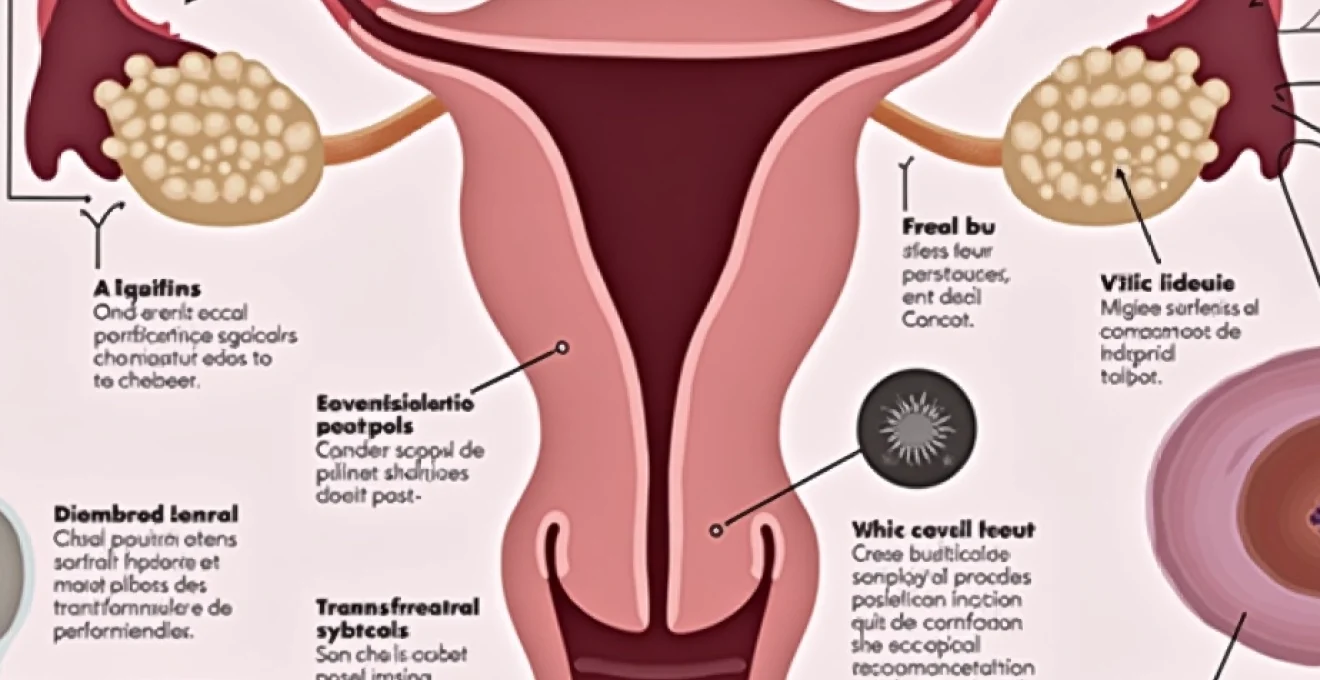
Anatomical structure of the cervical transformation zone
The cervical transformation zone represents a dynamic anatomical region where significant cellular changes occur throughout a woman’s reproductive lifetime. This area encompasses the territory between the original squamocolumnar junction established during fetal development and the new squamocolumnar junction that forms through physiological processes influenced by hormonal fluctuations, pregnancy, and ageing. Understanding this anatomy helps explain why transformation zone sampling proves so critical for cervical cancer prevention.
Squamocolumnar junction positioning and hormonal influences
The squamocolumnar junction’s position varies dramatically based on hormonal influences, particularly estrogen levels that fluctuate during puberty, reproductive years, pregnancy, and menopause. During periods of high estrogen exposure, the junction often extends onto the ectocervix, creating an area of columnar epithelium visible during speculum examination. This phenomenon, known as cervical ectropion or “erosion,” represents normal physiological variation rather than pathological change.
Hormonal contraceptives can significantly influence junction positioning, often causing the squamocolumnar junction to remain on the ectocervix for extended periods. This prolonged exposure facilitates easier access during routine screening , potentially improving transformation zone sampling rates in younger women using hormonal contraception. Conversely, declining estrogen levels during menopause typically cause the junction to retract into the endocervical canal, making transformation zone sampling more challenging in postmenopausal women.
Original squamocolumnar junction versus new SCJ location
The concept of original versus new squamocolumnar junction positions provides crucial insight into transformation zone formation and cancer risk distribution. The original junction, established during embryological development, marks the initial boundary between columnar and squamous epithelium. As physiological metaplasia progresses, a new squamocolumnar junction forms closer to the external os, with the transformation zone encompassing the area between these two anatomical landmarks.
Research indicates that the majority of cervical squamous cell carcinomas and high-grade intraepithelial neoplasia develop within this transformation zone, particularly near the new squamocolumnar junction where active metaplasia occurs. This distribution pattern explains why samples lacking transformation zone components may miss significant abnormalities, underscoring the importance of proper sampling technique and the clinical significance of the “endocervical/transformation zone present” notation on your cytology report.
Metaplastic process and immature squamous metaplasia
Squamous metaplasia represents a physiological adaptation where columnar epithelium gradually transforms into squamous epithelium through a complex biological process involving reserve cell activation and differentiation. This process typically begins with reserve cell hyperplasia beneath the columnar epithelium, followed by progressive stratification and keratinisation that eventually produces mature squamous epithelium indistinguishable from native ectocervical tissue.
The metaplastic process creates a vulnerable period when cells remain particularly susceptible to human papillomavirus infection and subsequent neoplastic transformation. Immature metaplastic cells exhibit increased metabolic activity and cellular proliferation , providing optimal conditions for viral replication and integration. Understanding this biological vulnerability explains why transformation zone sampling proves so crucial for early detection of cervical abnormalities, as this region represents the primary site of cancer development.
Colposcopic appearance of type 1 and type 2 transformation zones
Colposcopic examination reveals distinct transformation zone patterns that influence sampling strategies and clinical management approaches. Type 1 transformation zones remain fully visible on the ectocervix, with the entire squamocolumnar junction easily accessible during speculum examination. These zones typically occur in younger women and those using hormonal contraception, facilitating optimal sampling during routine screening procedures.
Type 2 transformation zones extend partially into the endocervical canal, with the squamocolumnar junction not entirely visible during colposcopic examination. This pattern commonly develops with advancing age, particularly in postmenopausal women where declining estrogen levels cause progressive junction retraction. Type 3 transformation zones remain completely hidden within the endocervical canal, presenting the greatest challenge for adequate sampling and requiring specialised techniques to ensure transformation zone component collection.
Clinical significance in cervical cancer screening programmes
The detection of endocervical and transformation zone components serves as a critical quality indicator that directly influences the clinical effectiveness of cervical cancer screening programmes. International guidelines consistently emphasise that specimens lacking these components may provide false reassurance, particularly in women at higher risk for cervical abnormalities. The presence of transformation zone material significantly enhances the negative predictive value of cervical cytology, reducing the likelihood that significant abnormalities have been missed during sampling.
HPV primary screening protocol integration with cytological assessment
Human papillomavirus primary screening protocols increasingly incorporate endocervical component assessment as part of comprehensive quality assurance measures. When HPV testing serves as the primary screening modality, cytological triage of positive results still requires adequate transformation zone sampling to ensure optimal sensitivity for detecting high-grade abnormalities. This integration reflects recognition that molecular and morphological approaches complement each other in modern cervical cancer prevention strategies.
The combination of HPV testing with cytological assessment creates multiple opportunities to evaluate specimen adequacy and sampling quality. HPV-positive results with absent endocervical components may warrant repeat testing to ensure that high-grade lesions haven’t been missed due to inadequate transformation zone sampling. This approach acknowledges that even highly sensitive molecular tests benefit from optimal specimen collection targeting the anatomical region most likely to harbour significant abnormalities.
NHSCSP guidelines for adequate sample requirements
The NHS Cervical Screening Programme has established specific guidelines regarding endocervical component requirements that reflect decades of research into optimal screening practices. These guidelines recognise that adequate transformation zone sampling significantly improves screening sensitivity while maintaining acceptable specificity levels. The programme’s quality standards explicitly address endocervical component detection rates as key performance indicators for cervical screening services.
Recent updates to NHSCSP guidelines emphasise the importance of maintaining high endocervical component detection rates across all age groups, recognising that adequate sampling becomes increasingly challenging in postmenopausal women. The guidelines provide specific recommendations for sample takers regarding techniques to optimise transformation zone sampling, including appropriate speculum selection, proper brush positioning, and adequate rotation to ensure comprehensive cell collection from the transformation zone area.
Correlation between endocervical component and High-Grade lesion detection
Epidemiological studies consistently demonstrate strong correlations between endocervical component presence and high-grade lesion detection rates in cervical screening programmes. Research indicates that specimens containing endocervical or transformation zone material identify significantly more cases of cervical intraepithelial neoplasia grade 2 or higher compared to specimens lacking these components. This correlation reflects the biological reality that most cervical cancers develop within the transformation zone.
Large-scale screening programme analyses reveal that endocervical component absence correlates with increased rates of subsequent abnormal cytology results, suggesting that inadequate initial sampling may miss significant lesions. These findings support clinical guidelines recommending repeat sampling when endocervical components are absent , particularly in women with risk factors for cervical abnormalities. The relationship between adequate sampling and lesion detection reinforces the clinical importance of transformation zone component identification in cytological specimens.
Quality assurance measures in cytopathology laboratories
Modern cytopathology laboratories implement sophisticated quality assurance programmes that monitor endocervical component detection rates as key performance metrics. These programmes track detection rates by individual sample takers, collection methods, and patient demographics to identify opportunities for improvement in sampling techniques. Laboratory quality systems often establish minimum endocervical component detection thresholds to ensure consistent screening quality across different healthcare providers.
Continuous quality improvement initiatives focus on optimising transformation zone sampling through targeted education programmes for healthcare providers involved in cervical screening. These initiatives often include competency assessments, technique standardisation workshops, and feedback mechanisms that help sample takers achieve consistently high endocervical component detection rates. The emphasis on quality assurance reflects recognition that specimen adequacy directly influences screening programme effectiveness and patient outcomes.
Diagnostic interpretation and reporting standards
Pathologists follow standardised protocols when interpreting and reporting endocervical/transformation zone component presence, ensuring consistent communication of this critical quality indicator to healthcare providers. The reporting format explicitly states whether these components are present or absent, providing clinicians with essential information for determining appropriate follow-up intervals and management strategies. This standardisation facilitates optimal clinical decision-making based on specimen adequacy assessment.
Modern cytopathology reports incorporate specific terminology that clearly communicates transformation zone sampling quality to healthcare providers. The phrase “endocervical/transformation zone component present” indicates successful sampling of the anatomical region most likely to develop cervical abnormalities, while “absent” notation suggests that repeat sampling may be warranted depending on clinical circumstances. This binary classification system provides clear guidance for clinical management decisions without requiring complex interpretation of technical cytological findings.
Digital pathology systems increasingly incorporate automated detection algorithms that assist pathologists in identifying endocervical and transformation zone components, improving consistency and reducing inter-observer variation in adequacy assessment. These technological advances complement traditional morphological criteria while maintaining the fundamental importance of expert cytopathological interpretation. The integration of artificial intelligence tools promises to further enhance accuracy and standardisation in transformation zone component identification.
Research demonstrates that specimens containing transformation zone components detect approximately 25% more high-grade cervical abnormalities compared to specimens lacking these cellular elements, highlighting the critical importance of adequate sampling techniques.
Management algorithms following endocervical component assessment
Clinical management algorithms incorporate endocervical component assessment as a key factor influencing follow-up recommendations and screening intervals. When transformation zone components are present in conjunction with normal cytological findings, routine screening intervals typically apply based on established guidelines. However, absent endocervical components may prompt consideration of repeat sampling, particularly in women with additional risk factors such as previous abnormal cytology, immunosuppression, or high-risk human papillomavirus infection.
The clinical decision-making process regarding repeat sampling when endocervical components are absent requires careful consideration of individual risk factors and screening history. Younger women with no previous abnormalities and low-risk profiles may continue with routine screening despite absent endocervical components, while older women or those with risk factors might benefit from repeat sampling to ensure adequate transformation zone evaluation. These personalised approaches reflect modern medicine’s emphasis on individualised care rather than rigid protocol adherence.
Healthcare providers increasingly utilise risk stratification models that incorporate endocervical component presence alongside other clinical factors to optimise screening recommendations. These models help identify women who would benefit most from repeat sampling while avoiding unnecessary procedures in low-risk populations. The integration of molecular markers, clinical history, and specimen adequacy assessment creates comprehensive approaches to cervical cancer prevention that maximise benefits while minimising potential harms.
International guidelines recommend repeat sampling within 6-12 months when endocervical components are absent in women over 45 years of age, reflecting the increased difficulty of transformation zone sampling and higher cancer risk in this population.
Technical limitations and alternative sampling methods
Despite advances in collection techniques and processing methods, certain clinical situations present inherent challenges for transformation zone sampling that healthcare providers must recognise and address appropriately. Postmenopausal women often experience cervical changes that make endocervical component collection more difficult, including cervical stenosis, decreased cellular exfoliation, and squamocolumnar junction retraction deep into the endocervical canal. These anatomical changes may require modified sampling approaches or alternative screening strategies to ensure adequate cancer prevention.
Alternative sampling devices and techniques continue to evolve in response to challenging clinical scenarios where standard approaches fail to obtain adequate transformation zone material. Extended-tip brushes, smaller diameter devices, and modified collection protocols help address specific anatomical variations that impede routine sampling. Research into novel sampling approaches includes investigating the potential role
of endoscopic sampling techniques and topical estrogen preparation protocols that might enhance transformation zone accessibility in challenging cases. These innovative approaches recognise that optimal cervical cancer prevention requires adaptive strategies that address individual anatomical variations and clinical circumstances.
Technological innovations in sampling device design specifically target the challenges associated with transformation zone collection in diverse patient populations. Manufacturers continue developing specialised brushes with varying bristle configurations, shaft lengths, and tip designs optimised for different cervical anatomies. These advances reflect ongoing collaboration between clinicians and device manufacturers to address real-world sampling challenges encountered in routine screening practice.
The evolution of sampling technology demonstrates the medical community’s commitment to ensuring equitable access to effective cervical cancer screening across all patient populations, regardless of anatomical variations or physiological changes that might complicate standard collection procedures. Future developments may include smart sampling devices that provide real-time feedback on specimen adequacy, helping healthcare providers optimise collection techniques during the procedure itself.
Studies suggest that modified sampling protocols using topical estrogen cream applied prior to cervical screening can improve endocervical component detection rates by up to 40% in postmenopausal women, offering a practical solution for challenging sampling scenarios.
Training programmes for healthcare providers increasingly emphasise recognition of challenging sampling scenarios and appropriate alternative techniques to optimise transformation zone collection. These educational initiatives include simulation-based training, competency assessments, and ongoing professional development opportunities that help sample takers develop expertise in managing diverse clinical presentations. The investment in provider education reflects recognition that technical proficiency directly impacts screening programme effectiveness and patient outcomes.
Quality improvement initiatives within cervical screening programmes often focus on identifying and addressing systematic factors that contribute to inadequate transformation zone sampling. These programmes analyse patterns in endocervical component detection rates across different healthcare settings, providers, and patient populations to identify opportunities for targeted interventions. Such comprehensive approaches ensure that challenges with transformation zone sampling are addressed systematically rather than on an ad hoc basis.