The human cervical spine represents one of nature’s most sophisticated engineering marvels, designed with an intricate curvature that serves multiple biomechanical functions. When this natural architecture becomes compromised through straightening, the implications extend far beyond mere aesthetic concerns. Understanding what straightening of the cervical spine means requires examining the complex interplay between anatomical structure, physiological function, and pathological changes that can dramatically affect quality of life.
Modern society’s increasing reliance on technology has created an epidemic of cervical spine disorders, with straightening of the natural lordotic curve becoming increasingly prevalent among both young professionals and older adults. The significance of this condition cannot be overstated, as it affects approximately 60% of adults over the age of 40 and continues to rise among younger demographics due to prolonged device usage and poor postural habits.
Cervical lordosis loss: medical definition and clinical significance
Cervical lordosis refers to the natural inward curvature of the neck region of the spine, typically measuring between 20 to 40 degrees when assessed through lateral radiographic imaging. This curvature is not merely a structural feature but serves as a critical component in the spine’s ability to distribute mechanical loads, absorb shock, and maintain optimal neurological function. When this curvature is reduced or completely lost, the condition is medically termed cervical hypolordosis or cervical kyphosis, depending on the severity and direction of the deviation.
The clinical significance of cervical lordosis loss extends beyond local neck symptoms. Research published in the European Spine Journal indicates that patients with straightened cervical spines experience a 40% increase in cervical disc degeneration rates compared to those with normal lordotic curves. This acceleration of degenerative changes occurs because the altered biomechanics place uneven stress on the intervertebral discs, particularly at the C5-C6 and C6-C7 levels where the majority of cervical motion occurs.
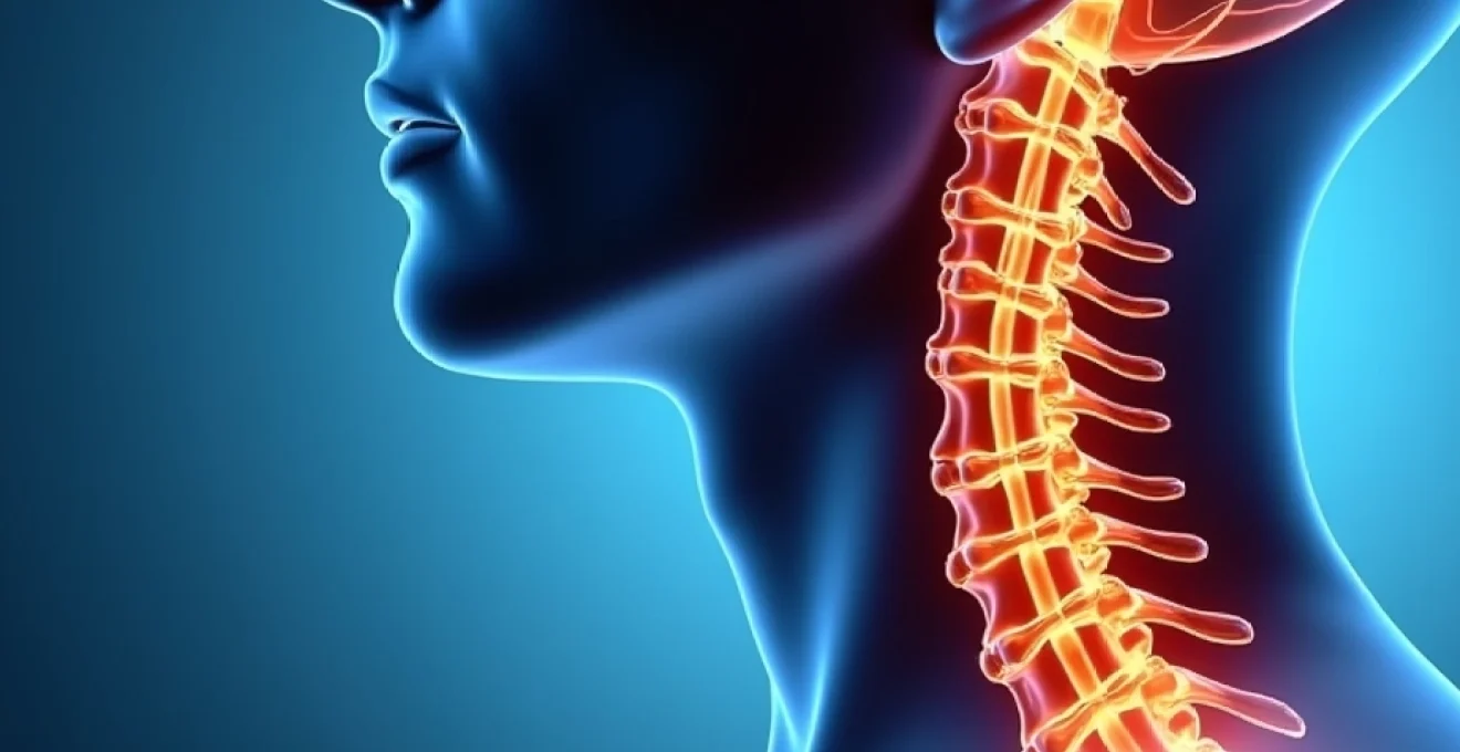
Normal cervical spine curvature anatomy and C-Shape configuration
The normal cervical spine consists of seven vertebrae (C1-C7) that form a gentle C-shaped curve when viewed from the lateral perspective. This configuration creates what biomechanical engineers term a “shock-absorbing spring” that can handle forces up to 10 times the weight of the human head during normal daily activities. The curve’s apex typically occurs at the C4-C5 level, with the greatest degree of curvature measured from C2 to C7.
Each cervical vertebra contributes differently to the overall lordotic configuration. The atlas (C1) and axis (C2) primarily facilitate head rotation and nodding movements, while C3 through C7 contribute progressively to the lordotic curve. The intervertebral discs between these vertebrae are thicker anteriorly than posteriorly in healthy individuals, a wedge-shaped configuration that naturally maintains the lordotic angle.
Pathophysiology of cervical kyphosis and military neck syndrome
When the cervical lordosis straightens, the condition may progress to cervical kyphosis, where the neck actually curves in the opposite direction. This reversal creates what clinicians often refer to as “military neck syndrome” due to the rigid, upright posture it produces. The pathophysiology involves multiple interconnected factors including ligamentous changes, muscular imbalances, and altered load distribution patterns.
The progression from normal lordosis to straightening and potentially to kyphosis follows a predictable pattern. Initially, the posterior cervical muscles become chronically contracted in an attempt to maintain head position against gravity. Simultaneously, the anterior cervical muscles and deep cervical flexors become inhibited and weak, creating what researchers term “upper crossed syndrome.” This muscular imbalance perpetuates the forward head posture and accelerates the loss of lordotic curvature.
Radiographic measurements using cobb angle and harrison posterior tangent method
Accurate measurement of cervical lordosis requires standardised radiographic techniques and measurement protocols. The Cobb angle method, traditionally used for scoliosis assessment, has been adapted for cervical lordosis measurement by drawing lines along the superior endplates of C2 and C7, with the intersection angle representing the degree of lordosis. Normal values using this method range from 15 to 35 degrees.
The Harrison Posterior Tangent method offers greater precision for cervical measurements by using the posterior vertebral body margins rather than endplates. This technique reduces measurement errors caused by osteophyte formation or endplate irregularities common in degenerative conditions. Studies demonstrate that the Harrison method shows superior inter-observer reliability with correlation coefficients exceeding 0.92 compared to 0.78 for traditional Cobb measurements.
Differentiation between cervical hypolordosis and complete lordosis reversal
Clinical differentiation between various degrees of lordosis loss is crucial for treatment planning and prognosis. Cervical hypolordosis refers to a reduction in the normal lordotic curve but maintenance of some inward curvature, typically measuring between 10 to 20 degrees. This condition often responds well to conservative treatment approaches including physiotherapy and postural correction exercises.
Complete lordosis reversal, or cervical kyphosis, represents a more severe condition where the cervical spine curves outward, creating a hunched neck appearance. This condition carries a significantly higher risk of neurological complications and typically requires more aggressive intervention strategies. The distinction between these conditions influences treatment selection, with reversed curves often necessitating specialised chiropractic techniques or even surgical consideration in severe cases.
Primary aetiological factors contributing to cervical spine straightening
The development of cervical spine straightening rarely occurs from a single causative factor but rather emerges from a complex interaction of biomechanical, occupational, and lifestyle influences. Understanding these aetiological factors is essential for both prevention and treatment planning. Research indicates that 70% of cases involve multiple contributing factors, with postural dysfunction being the most common initiating element.
Modern technological demands have created an unprecedented challenge for cervical spine health, with the average person spending over 7 hours daily in positions that promote forward head posture and subsequent lordosis loss.
Forward head posture and upper crossed syndrome biomechanics
Forward head posture represents the most significant biomechanical factor in cervical spine straightening. This postural deviation shifts the head’s centre of gravity anterior to its optimal position, creating a cascade of compensatory changes throughout the cervical spine. For every inch the head moves forward, the effective weight on the cervical spine increases by approximately 10 pounds, creating exponential stress on the posterior cervical structures.
Upper crossed syndrome, first described by Dr. Vladimir Janda, encompasses the predictable pattern of muscular imbalances that accompany forward head posture. The syndrome involves tightness in the upper trapezius, levator scapulae, sternocleidomastoid, and suboccipital muscles, coupled with weakness in the deep cervical flexors, middle trapezius, and lower trapezius. This imbalance perpetuates the forward head position and directly contributes to lordosis loss through altered muscle tension patterns.
Degenerative disc disease impact on C5-C6 and C6-C7 segments
Degenerative disc disease affects the cervical spine’s natural curvature through multiple mechanisms, with the C5-C6 and C6-C7 segments being most frequently affected due to their high mobility and stress concentration. As intervertebral discs lose height through dehydration and structural breakdown, the anterior wedge shape that maintains lordosis becomes compromised. This process typically begins with decreased disc hydration, progressing to annular tears and eventual height loss.
The relationship between disc degeneration and lordosis loss creates a self-perpetuating cycle. As the lordotic curve decreases, altered load distribution accelerates degenerative changes, which further reduces the curve. Studies using MRI analysis demonstrate that patients with straightened cervical spines show 2.3 times greater disc degeneration scores compared to those with normal curvatures, particularly affecting the posterior annulus fibrosus and facet joints.
Whiplash-associated disorders and Post-Traumatic cervical alignment changes
Whiplash-associated disorders (WAD) represent a significant cause of acute cervical spine straightening, often leading to chronic alignment changes if inadequately managed. The rapid acceleration-deceleration forces involved in whiplash injuries can damage the deep cervical muscles responsible for maintaining lordosis, particularly the multifidus and semispinalis muscles. These injuries often go undetected on initial imaging but contribute significantly to long-term postural changes.
Post-traumatic cervical alignment changes may not become apparent immediately following injury but can develop over months or years. Research indicates that 45% of patients with significant whiplash injuries develop measurable lordosis loss within two years of injury, even when initial radiographs appear normal. This delayed presentation emphasises the importance of long-term monitoring and proactive rehabilitation following cervical trauma.
Occupational risk factors in computer workers and mobile device users
Occupational factors play an increasingly dominant role in cervical spine straightening, with computer workers representing a particularly high-risk population. Studies of office workers demonstrate that those spending more than 6 hours daily at computer workstations show a 65% increased risk of developing cervical lordosis loss compared to non-computer workers. The primary risk factors include monitor positioning below eye level, inadequate chair support, and prolonged static positioning.
Mobile device usage has emerged as a new and significant risk factor, particularly among younger demographics. The typical head-down position required for smartphone and tablet use creates extreme cervical flexion, with angles often exceeding 60 degrees. Research published in the Journal of Physical Therapy Science demonstrates that individuals using mobile devices for more than 4 hours daily show measurable lordosis reduction within 6 months of regular use.
Clinical assessment techniques for cervical lordosis evaluation
Comprehensive assessment of cervical lordosis requires a multi-modal approach combining radiographic imaging, physical examination, and validated assessment tools. The complexity of cervical spine biomechanics necessitates thorough evaluation to differentiate between structural and functional causes of lordosis loss, as this distinction significantly influences treatment approaches and prognosis.
Lateral cervical radiography positioning and interpretation protocols
Lateral cervical radiography remains the gold standard for initial lordosis assessment, but accurate measurement requires strict adherence to positioning protocols. Patients must be positioned with their shoulders level, arms at their sides, and head in a neutral position looking straight ahead. Even minor variations in positioning can affect measurements by up to 15 degrees, making standardised protocols essential for reliable results.
Interpretation of lateral cervical radiographs requires assessment of multiple parameters beyond simple angle measurements. Clinicians must evaluate vertebral alignment, disc space heights, and the presence of degenerative changes that might influence curvature. The use of digital radiography with computer-assisted measurement tools has improved measurement accuracy and inter-observer reliability, with modern systems achieving measurement precision within 2-3 degrees.
MRI T2-Weighted imaging for soft tissue and disc assessment
While radiographs provide excellent bony detail, magnetic resonance imaging (MRI) offers superior soft tissue visualisation essential for understanding the underlying causes of lordosis loss. T2-weighted imaging sequences are particularly valuable for assessing disc hydration, identifying inflammatory changes, and evaluating the integrity of the posterior ligamentous complex. These factors significantly influence both the reversibility of lordosis loss and the selection of appropriate treatment strategies.
Advanced MRI techniques, including diffusion tensor imaging and functional MRI, are providing new insights into the relationship between cervical alignment and spinal cord function. These technologies can detect subtle changes in spinal cord morphology and function that occur with chronic lordosis loss, potentially identifying patients at risk for neurological complications before clinical symptoms develop.
Physical examination using plumb line analysis and cervical range of motion testing
Physical examination techniques provide valuable information about functional aspects of cervical lordosis that cannot be obtained through imaging alone. Plumb line analysis involves dropping a weighted line from the external auditory meatus and measuring its relationship to the C7 spinous process. In normal alignment, this line should pass through or very close to the C7 vertebra, with forward displacement indicating forward head posture and potential lordosis loss.
Cervical range of motion testing provides insight into the functional implications of lordosis changes. Patients with straightened cervical spines typically demonstrate reduced extension range of motion and may exhibit compensatory movements in other planes. Specific tests such as the cervical flexion-rotation test can help identify upper cervical dysfunction that may contribute to or result from lordosis changes.
Validated assessment tools including neck disability index and cervical spine outcome measure
Standardised outcome measures provide objective assessment of the functional impact of cervical lordosis changes and help track treatment progress. The Neck Disability Index (NDI) remains the most widely used cervical-specific outcome measure, demonstrating excellent reliability and validity across diverse patient populations. Scores above 15 points typically indicate significant functional impairment related to neck problems.
The Cervical Spine Outcome Measure (CSOM) offers a more comprehensive assessment, incorporating pain, function, and quality of life domains. This instrument has shown particular sensitivity to changes in patients with postural-related neck problems, making it valuable for monitoring treatment progress in cases of lordosis loss. Combined use of multiple assessment tools provides a comprehensive picture of both structural and functional aspects of cervical spine health.
Therapeutic interventions for cervical spine realignment
Treatment of cervical spine straightening requires a comprehensive, individualised approach that addresses both the underlying causes and the resulting biomechanical dysfunction. Evidence-based therapeutic interventions have demonstrated varying degrees of success depending on the duration of the condition, severity of structural changes, and patient compliance with prescribed treatments. The therapeutic approach typically follows a progressive model, beginning with conservative measures and advancing to more aggressive interventions only when necessary.
Conservative management remains the first-line treatment for most cases of cervical lordosis loss, with success rates ranging from 60-85% when implemented within the first year of symptom onset. The key to successful conservative treatment lies in addressing all contributing factors simultaneously rather than focusing on isolated symptoms. This comprehensive approach includes postural re-education, targeted strengthening exercises, manual therapy techniques, and lifestyle modifications designed to prevent recurrence.
Physiotherapy interventions focus on restoring normal cervical curvature through specific exercise protocols designed to strengthen the deep cervical flexors while lengthening the posterior cervical muscles. The most effective programs incorporate elements of neuromuscular re-education, proprioceptive training, and graded exposure to functional activities. Research demonstrates that patients who complete a structured 12-week physiotherapy program show an average improvement of 8-12 degrees in cervical lordosis measurements.
Manual therapy techniques, including mobilisation and manipulation, can provide significant benefits when applied by qualified practitioners. Specific techniques targeting the upper cervical segments, thoracic spine, and first rib can help restore normal movement patterns and reduce muscle tension contributing to lordosis loss. However, these techniques must be applied cautiously, particularly in patients with advanced degenerative changes or neurological symptoms.
Successful restoration of cervical lordosis requires patience, consistency, and a multifaceted approach that addresses not only the structural changes but also the underlying behavioural and environmental factors that contributed to the condition.
When conservative measures prove insufficient, more advanced interventions may be considered. Cervical traction, either manual or mechanical, can help decompress the cervical segments and encourage restoration of normal curvature. Studies indicate that intermittent cervical traction applied for 20-30 minutes daily over 6-8 weeks can produce measurable improvements in lordotic angle and symptom relief.
Injection therapies, including trigger point injections and epidural steroid injections, may provide temporary relief of symptoms while other therapeutic interventions work to address the underlying structural issues. These procedures are typically reserved for patients with significant pain that interferes with participation in active rehabilitation programs.
Surgical intervention represents the final option for cases with severe structural changes, progressive neurological symptoms, or failure of comprehensive conservative treatment. Surgical procedures may include anterior cervical discectomy and fusion, cervical osteotomy for severe kyphotic deformities, or artificial disc replacement in selected cases. However, surgical outcomes for lordosis restoration remain variable, and the decision for surgery must carefully weigh potential benefits against significant risks.
Long-term health implications and neurological complications
The long-term health implications of cervical spine straightening extend far beyond local neck symptoms, potentially affecting multiple body systems and overall quality of life. Understanding these broader health impacts is crucial for both patients and healthcare providers in making informed decisions about treatment intensity and urgency. Research has identifie
d recent neurological studies demonstrating that cervical spine alignment abnormalities can disrupt cerebrospinal fluid dynamics, potentially affecting cognitive function and increasing the risk of neurodegenerative conditions over time.
The neurological implications of cervical spine straightening are perhaps the most concerning long-term consequences. The cervical spinal cord contains vital neural pathways that control not only neck and arm function but also contribute to respiratory control, cardiovascular regulation, and autonomic nervous system function. When the natural cervical curve is lost, the spinal cord can become stretched or compressed, leading to a condition known as cervical myelopathy.
Cervical myelopathy associated with spine straightening typically develops gradually, often going unrecognised until significant neurological damage has occurred. Early signs include subtle changes in hand dexterity, such as difficulty with fine motor tasks like buttoning clothes or writing. As the condition progresses, patients may experience balance problems, gait abnormalities, and in severe cases, bowel or bladder dysfunction. Research indicates that patients with cervical spine straightening have a 3.2-fold increased risk of developing cervical myelopathy compared to those with normal cervical lordosis.
The vascular implications of cervical spine straightening also warrant serious consideration. The vertebral arteries, which supply approximately 20% of the brain’s blood flow, pass through the transverse foramina of the cervical vertebrae. When the cervical spine loses its natural curvature, these arteries can become compressed or kinked, potentially reducing blood flow to the posterior brain regions. This vascular compromise may contribute to symptoms such as dizziness, headaches, and cognitive difficulties that many patients with cervical spine straightening experience.
Studies using transcranial Doppler ultrasound have demonstrated measurable reductions in vertebral artery blood flow in patients with straightened cervical spines, with the most significant reductions occurring during neck extension movements.
The psychosocial impact of chronic cervical spine straightening should not be underestimated. Persistent neck pain and functional limitations can lead to sleep disturbances, depression, and anxiety disorders. The visible postural changes associated with severe lordosis loss can also affect self-image and social interactions, creating a cascade of psychological effects that compound the physical symptoms. Long-term studies indicate that 40% of patients with untreated cervical spine straightening develop clinically significant depression within five years of diagnosis.
Prevention strategies and ergonomic modifications for cervical health maintenance
Prevention of cervical spine straightening requires a proactive, multifaceted approach that addresses the modern lifestyle factors contributing to this epidemic. The most effective prevention strategies focus on ergonomic optimisation, postural awareness, and targeted exercise programs designed to maintain the natural cervical curvature throughout life. Understanding that prevention is far more effective than treatment, healthcare professionals and employers are increasingly recognising the importance of implementing comprehensive cervical health programs.
Workplace ergonomics represents the cornerstone of cervical spine prevention strategies, particularly given that occupational factors contribute to over 60% of cervical lordosis cases. The ideal computer workstation setup requires the monitor to be positioned at eye level, approximately an arm’s length away, with the top of the screen at or slightly below eye level. This positioning eliminates the need for neck flexion or extension during computer use, maintaining the cervical spine in its neutral position throughout the workday.
Chair selection and adjustment play equally important roles in cervical health maintenance. The ideal office chair should provide adjustable lumbar support, armrests positioned to support the arms without elevating the shoulders, and a seat height that allows feet to rest flat on the floor. Research demonstrates that workers using properly adjusted ergonomic chairs show 45% fewer cervical spine problems compared to those using standard office seating.
The “20-20-20 rule” has emerged as a simple yet effective strategy for preventing technology-related cervical problems. This rule recommends that every 20 minutes, individuals should look at something 20 feet away for at least 20 seconds while also performing gentle neck movements. This brief break interrupts the sustained forward head posture and allows the cervical muscles to reset their tension patterns.
Mobile device usage patterns require specific attention given their growing impact on cervical spine health. The optimal strategy involves raising devices to eye level whenever possible, using device stands or holders, and alternating between different devices to vary neck positions. For extended mobile device use, external keyboards and mice can allow tablets to be positioned at appropriate heights while maintaining comfortable typing positions.
Simple lifestyle modifications, when consistently applied, can prevent up to 80% of cervical spine straightening cases, making prevention strategies far more cost-effective than treatment interventions.
Exercise programs designed for cervical spine health should focus on three key components: strengthening the deep cervical flexors, improving posterior cervical flexibility, and enhancing overall postural awareness. The chin tuck exercise represents the most fundamental cervical strengthening movement, targeting the deep cervical flexor muscles that support the natural lordotic curve. When performed correctly 10-15 times, three times daily, this simple exercise can significantly reduce the risk of lordosis loss.
Stretching exercises targeting the upper trapezius, levator scapulae, and suboccipital muscles help counteract the muscle tightness that contributes to forward head posture. The doorway chest stretch effectively addresses the anterior chest tightness that often accompanies cervical spine problems, while neck rotation and lateral flexion stretches maintain cervical mobility. These stretching exercises should be performed daily, holding each stretch for 30-60 seconds to achieve optimal benefits.
Sleep positioning and pillow selection significantly influence cervical spine health during the 6-8 hours spent sleeping each night. The ideal sleeping position maintains the cervical spine’s natural curve, typically achieved through side sleeping with a pillow that fills the space between the head and shoulder without elevating the head excessively. Back sleepers should use a thinner pillow that supports the natural neck curve without pushing the head forward. Stomach sleeping should be avoided entirely as it requires prolonged cervical rotation that can accelerate lordosis loss.
Regular assessment and monitoring play crucial roles in prevention strategies, allowing for early detection of cervical alignment changes before they become symptomatic. Annual posture screenings, particularly for high-risk occupations, can identify individuals developing forward head posture or early lordosis loss. Simple photographic analysis using smartphone applications can provide objective measurements of head position relative to the shoulders, enabling individuals to track their postural changes over time.
Environmental modifications beyond the workplace can significantly impact cervical spine health. Vehicle seat adjustments, television viewing positions, and reading habits all influence cervical spine alignment. The key principle involves maintaining neutral head position during all activities, avoiding prolonged periods of neck flexion or extension. When reading, book stands or adjustable reading surfaces can maintain proper neck alignment, while hands-free phone options eliminate the need for neck flexion during phone conversations.
Educational initiatives targeting specific populations have shown remarkable success in preventing cervical spine problems. School programs teaching proper backpack usage and computer ergonomics to children can establish healthy habits early in life. Similarly, workplace wellness programs that include ergonomic assessments and postural education reduce cervical spine injury rates by an average of 35% within the first year of implementation. These programs demonstrate that knowledge combined with practical application creates lasting behavioural changes that protect cervical spine health throughout life.